Introduction
You might work through this system over several weeks, days or hours, but to enhance your learning and enjoyment make sure you break it up into bite-size chunks.
Here are the sections of the Nervous System…
Make notes as you study each section, and interact fully with the activities – watch the animations and complete the quizzes.
Take a break at the end of each section– resting your eyes from the computer screen, getting some fresh air or taking a coffee break will improve your ability to focus on your study and take in information.
Give yourself time to think about what you have learned, and time to absorb and understand it.
 UHI / CC0
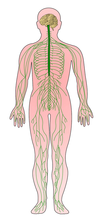
The nervous system
Together with the endocrine system, the nervous system is responsible for maintaining homeostasis within the body - maintaining all systems within their normal physiological limits in order to maintain health.
The nervous system does this by sending electrical signals called action potentials to tissues and organs in the body.
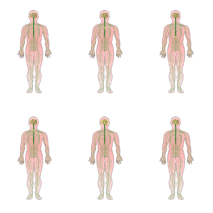
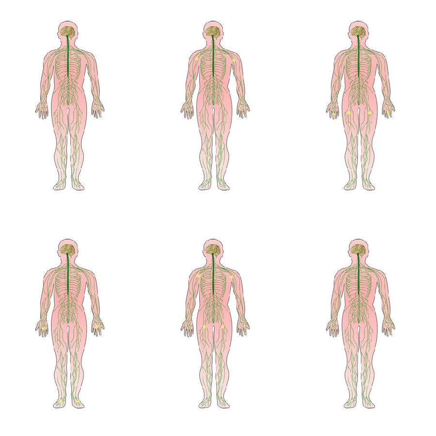
The nervous system has three basic functions:
Sensory function:
We obtain information
Integrative function:
We decide what to do with the onformation
Motor function:
We create an action

UHI / CC0
(Click image to toggle animation on/off)
UHI / CC0
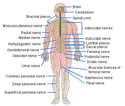
Brachial plexus
Musculocutaneous nerve
Radial nerve
Median nerve
Lliohypogastric nerve
Genitofemoral nerve
Obturator nerve
Ulnar nerve
Common peroneal nerve
Deep peroneal nerve
Superficial peroneal nerve
Brain
Cerebellum
Spinal cord
Intercostal nerves
Subcostal nerve
Lumbar plexus
Sacral plexus
Femoral nerve
Pudendal nerve
Sciatic nerve
Muscular branches of femoral nerve
Saphenous nerve
Tibial nerve
How the Nervous System Works
Sensory function
The nervous system senses changes inside and outside of the body, such as feeling sensations on the skin or changes in the chemicals in the blood.
Integrative function
We analyze the information received, store some information, and make decisions. For example, we may pull away from a hot surface quickly (motor function) due to the pain felt upon touching it (sensory feedback), and remember that the surface is hot.
Motor function
The nervous system may initiate muscular contraction or glandular secretion in response to stimuli from receptors all over the body.
The organisation of the nervous system
The nervous system has two main divisions: the Central Nervous System (CNS) and the Peripheral Nervous System (PNS).
UHI / CC0
(Click image to toggle animation on/off)
UHI / CC0
(Click image to toggle animation on/off)
The central nervous system consists of the brain and spinal cord. The peripheral nervous system consists of the nerves branching off from the brain and spinal cord.
Information is carried along nerves between the central nervous system and the peripheral nervous system.
Nerves in the peripheral nervous system collect information from the peripheral (outer) areas of the body via large numbers of sensory receptors and convey this information via nerves back to the central nervous system for processing.
The central nervous system
The central nervous system forms and stores memories, deals with incoming sensory information, generates emotions and thoughts and is responsible for stimulating most of the muscular contractions and glandular secretions that take place.
In the central nervous system (CNS), the brain and spinal cord form a central ‘axis’ in the body from which nerves run outwards to all parts of the body, connecting the central nervous system to sensory receptors, glands and muscles.
Nerves arise in pairs (left and right sides) from the brain (cranial nerves) and from the spine (spinal nerves). There are 12 pairs of cranial nerves and 31 pairs of spinal nerves.

UHI / CC0
(Click image to toggle animation on/off)
Sending information around the body
Nerve cells (neurones) in the PNS conduct nerve impulses from sensory receptors in various parts of the body to the CNS. These neurones are called afferent or sensory neurones. Sensory nerves carry information to the CNS along an afferent nerve.
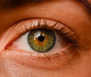
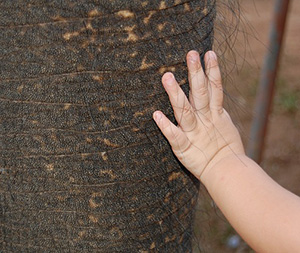
We pick up sensory information from our eyes, ears, skin, tongue, nose and muscles.
Motor or efferent neurones originate within the CNS and conduct nervous impulses from the CNS to muscles and glands. Motor nerves send information from the CNS to the muscles via an efferent nerve.
The peripheral nervous system
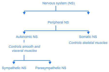
The peripheral nervous system is sub-divided into the somatic nervous system (voluntary) and the autonomic nervous system (involuntary).
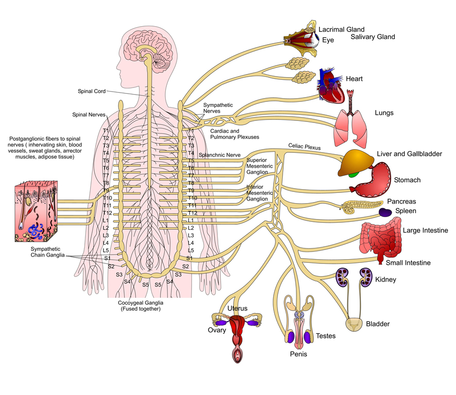
Sympathetic nervous system
UHI / CC0
- Spinal cord
- Spinal nerves
- Postganglionic fibres to spinal nerves (innervating skin, blood vessels, sweat glands, arrector muscles, adipose tissue)
- Sympathetic chain ganglia
- Coccygeal ganglia (fused together)
- Sympathetic nerves
- Cardiac and pulmonary plexuses
- Splanchnic nerve
- Celiac plexus
- Superior mesenteric ganglion
- Inferior mesenteric ganglion
- Lacrimal gland
- Eye
- Salivary gland
- Heart
- Lungs
- Liver and Gallbladder
- Stomach
- Pancreas
- Spleen
- Large intestine
- Small intestine
- Kidney
- Bladder
- Testes
- Penis
- Uterus
- Ovary
Peripheral nervous system
UHI / CC0
Flow chart:
Nervous system
Peripheral nervous system
Somatic NS - Controls skeletal muscles
Autonomic NS - Controls smooth and visceral muscles
Sympathetic NS
Parasympathetic NS
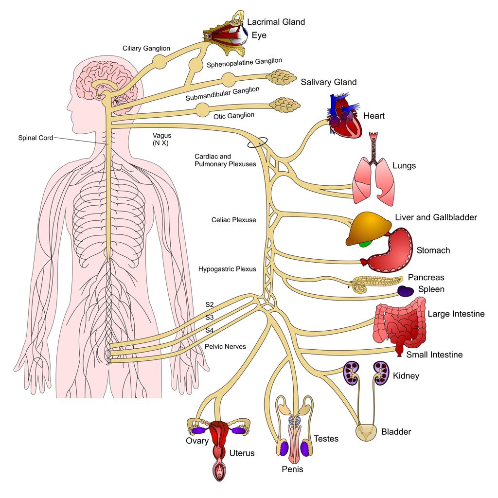
Parasympathetic nervous system
UHI / CC0
- Spinal cord
- Ciliary ganglion
- Lacrimal gland
- Eye
- Sphenopalatine ganglion
- Submandibular ganglion
- Otic ganglion
- Vagus (N X)
- Cardiac and pulmonary plexuses
- Celiac plexus
- Hypogastric plexus
- S2, S3, S4 pelvic nerves
- Salivary gland
- Heart
- Lungs
- Liver and Gallbladder
- Stomach
- Pancreas
- Spleen
- Large intestine
- Small intestine
- Kidney
- Bladder
- Testes
- Penis
- Uterus
- Ovary
The autonomic (involuntary) nervous system consists of sensory (afferent) neurones that convey information from sensory receptors, and efferent (motor) nerve cells which convey messages to glands, cardiac and smooth muscle such as the blood vessels. We do not consciously control these functions, therefore this is involuntary homeostasis, creating changes in rate and strength of heartbeat, vasoconstriction and vasodilation of blood vessels, or stimulation and depression of glandular secretions.
The somatic (voluntary) nervous system consists of sensory (afferent) neurones that convey information from sensory receptors around the body, and motor (efferent) nerve cells which convey messages on to skeletal muscles. Movements of the skeletal muscles can be consciously controlled, therefore this is a voluntary movement.
The autonomic nervous system
The Autonomic Nervous System has two divisions: the sympathetic and parasympathetic divisions.
Sympathetic
- Prepares the body for emergency situations
- Involves energy expenditure
- At rest, it counteracts (balances) the PNS
- During stress, it dominates the PNS
- Exercise, fear or anger stimulate this system
Parasympathetic
- Balances stimulation from the sympathetic nervous system
- Regulates activities that conserve and restore energy during rest
- The PNS governs responses such as salivation, tear flow, urination, digestion and defecation.
The sympathetic division generally prepares the body for emergency situations and usually involves energy expenditure. At rest, the sympathetic nervous system counteracts (balances) the parasympathetic nervous system, but during stress, the sympathetic system dominates. Exercise and emotions such as fear or anger stimulate the sympathetic nervous system. The parasympathetic nervous system regulates activities that conserve and restore body energy during rest. Parasympathetic impulses to the digestive system dominate sympathetic impulses, allowing the digestion of food to take place.
Quiz
The brain and spinal cord
Brain
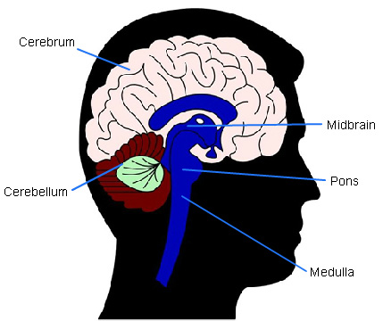
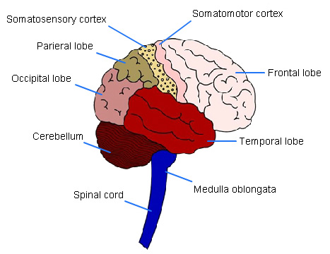
The brain is made up of billions of neurones and neuroglia (non-excitable) cells. It consists of four major parts:
The brain stem is continuous with the spinal cord and is made up of the medulla oblongata, pons and midbrain.
The cerebellum is at the back of the brain, behind the brain stem.
Above the brain stem is the diencephalon, which consists of the thalamus, hypothalamus, pineal gland and epithalamus.
The cerebrum (made up of two cerebral hemispheres) sits on top of the other parts of the brain and occupies most of the cranium.
UHI /CC0
- Somatosensory cortex
- Somatomotor cortex
- Parieral lobe
- Occipital lobe
- Frontal lobe
- Cerebellum
- Temporal lobe
- Spinal cord
- Medulla oblongata
Spinal cord
The spinal cord runs from the brain stem down through the centre of the vertebral column and is protected by the vertebrae. It forms the communication pathway between the brain and the body and allows nervous impulses to travel from the peripheral nervous system (the organs and limbs) to the central nervous system (brain and spinal cord) via the nerves which extrude from between the vertebrae.
It allows signals to pass down from the brain to control body function and enables messages to travel up to inform the brain of what is happening in the body.
This diagram shows a cross-section of the spinal cord running down the spine, and shows how nervous impulses are sent from a receptor gland or muscle, through the spinal cord and a motor impulse sent on to skeletal muscle via a motor neuron.
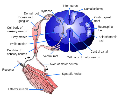
UHI CC0
- Interneuron
- Dorsal column
- Corticospinal tract
- Rubrospinal tract
- Spinothalamic tract
- Central canal
- Cell body of motor neuron
- Ventral root
- Axon of motor neuron
- Synaptic knobs
- Effector muscle
- Receptor
- Synapse
- Dorsal root
- Dorsal root ganglion
- Cell body of sensory neuron
- Grey matter
- White matter
- Dendrite of sensory neuron
Controlling the right and left sides of the body
Most motor and sensory tracts cross over from right to left or left to right as they pass through the medulla of the brain. The axons crossing over in the pyramids of the medulla is called decussation.
When a muscle is moved, a conscious decision originates in the cerebral cortex of the brain and impulses travel down from the right side of the brain across the medulla, go down the spinal cord, and result in moving the muscle in the left leg. Nervous impulses can pass very rapidly from the brain via the spinal cord to a muscle, enabling a rapid response and a decision via a voluntary process.
The right cerebral cortex controls the left side of the body.
The left cerebral cortex controls the right side of the body.
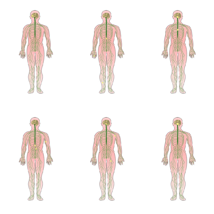
UHI / CC0
(Click image to toggle animation on/off)
Nerves and neurones
The basic unit of the nervous system is a cell called a neurone (or neuron). A neurone consists of a cell body, a dendrite and an axon. There are 3 main types of neuron: motor, sensory and connector (relay).
Motor nerves contain motor fibres which move muscles Sensory nerves contain sensory fibres to carry sensory impulses
Mixed nerves contain both motor and sensory fibres e.g. spinal nerves.
Some neurones are microscopic but others may have axons of over one metre in length. Bundles of neurones are collectively known as nerves and these are arranged in bundles (tracts) in the central nervous system. In the peripheral nervous system, they are arranged in clusters called ganglia.
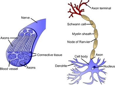
UHI / CC0
- Nerve
- Axons
- Blood vessel
- Connective tissue
- Axon terminal
- Schwann cell
- Myelin sheath
- Node of Ranvier
- Cell body
- Axon
- Dendrite
- Nucleus
The Structure of a Neurone
Specialized cells called Schwann cells form a protein-lipid layer around the axon called the myelin sheath. This provides protection and insulation for the electrical nervous impulse and increases the speed of the nervous impulse.
This layer is interrupted at intervals by narrow gaps called the Nodes of Ranvier where the axon membrane is exposed to the surrounding tissue fluid (containing sodium ions), making action potentials possible only at these gaps. The action potential (nervous impulse) ‘jumps’ from node to node along the axon allowing impulses to travel along an axon very quickly. Some nerves, however, do not contain myelin and are said to be un- myelinated.
Neurons are positioned end to end but do not touch – the gap in between each neurone is called a synaptic cleft or synapse. The axon terminal of one neuron lies next to the dendrite of another. The axon terminal ends in a bulbous structure called the synaptic knob, and this contains vesicles that hold chemicals called neurotransmitters.
Neurotransmitters can continue the nervous impulse on to the next neurone by travelling across the synaptic cleft, through the post-synaptic membrane of a dendrite in the neighbouring neurone and stimulating an action potential along the next neuron.
Synapses are essential along the nerve fibres as they control the passage of information from one neurone to the next. Impulses move from a pre-synaptic neurone (before the synapse), across a synaptic cleft and onto the post-synaptic neuron.
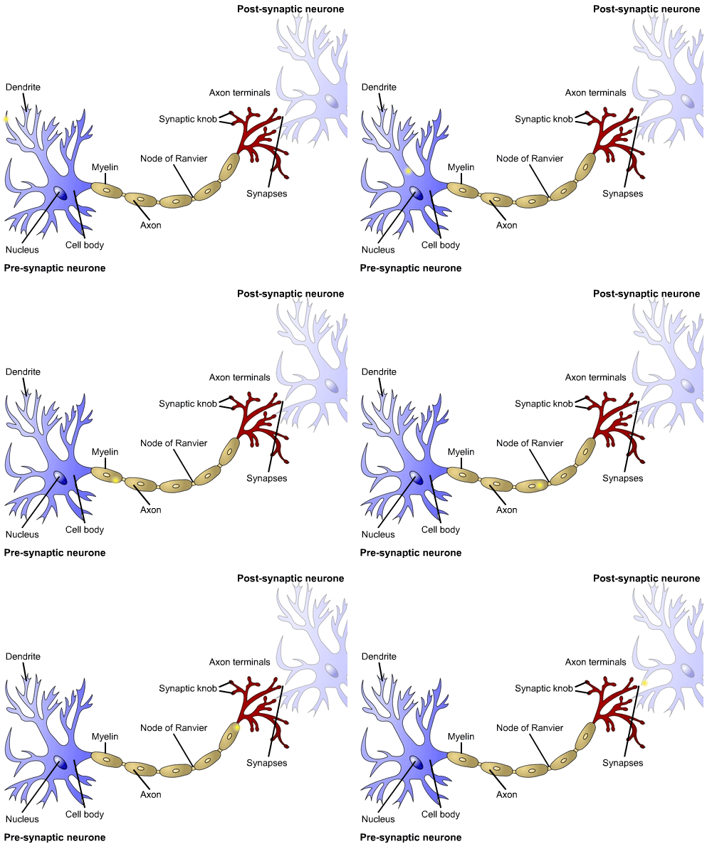
UHI / CC0
(Click image to toggle animation on/off)
Pre-synaptic neurone:
- Axon terminals
- Synapses
- Synaptic knob
- Myelin
- Node of Ranvier
- Cell body
- Axon
- Dendrite
- Nucleus
Quiz
The nervous impulse
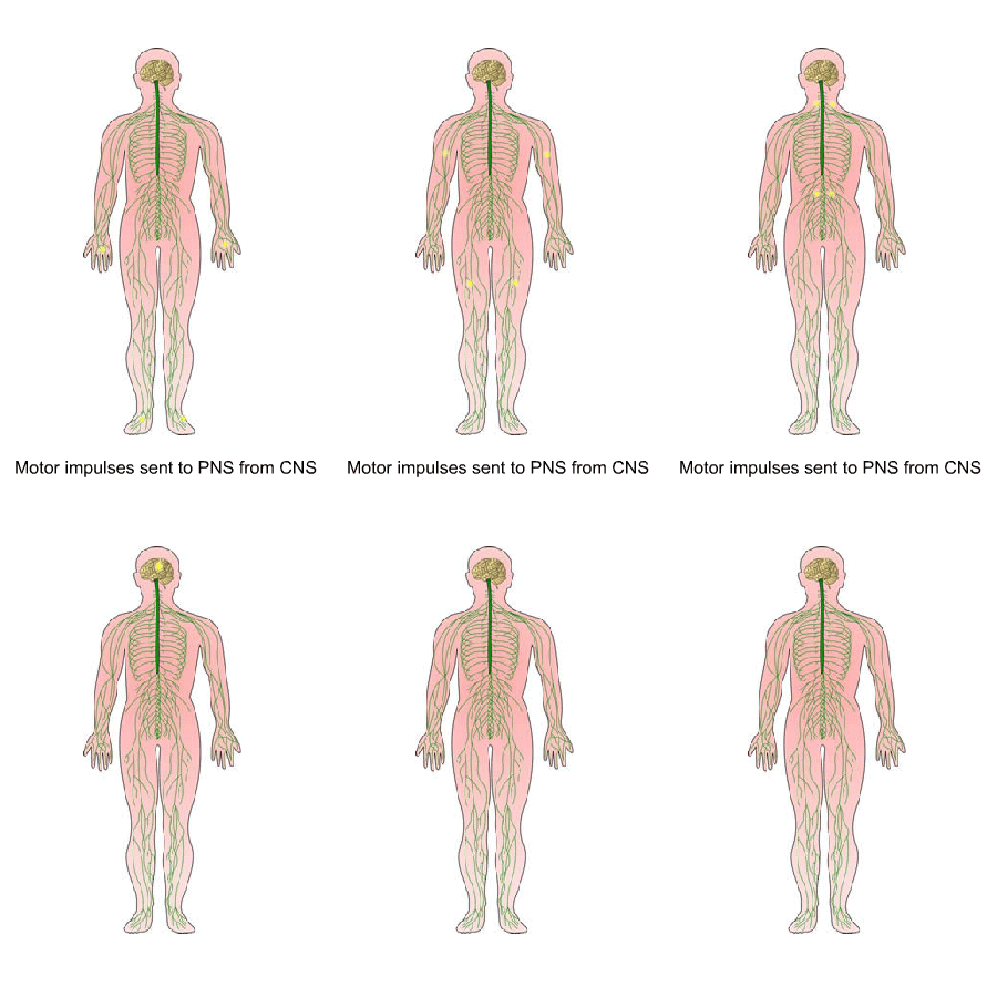
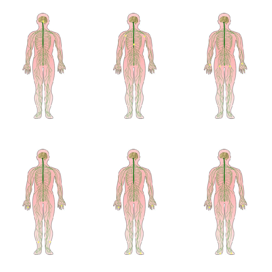
Whatever the type of stimulus or response, and whether the impulse is in the central or peripheral, autonomic or somatic nervous systems, the chain of events in the nervous system is always the same.
- A stimulus triggers the receptors
- A message is sent to the brain or spinal cord via the sensory (afferent) neurons
- The information is received in the CNS and a response is formulated.
- A message is sent from the CNS to the PNS (gland or muscle) via the efferent (motor) neurons (with the help of connector neurons).
- The muscle(s) or gland(s) (effector) respond
If the nervous impulse is stopped at some point along the nerve fibre by an inhibitory neurotransmitter or a drug (e.g. a pain killer) at the axon terminal (end of the nerve fibre), the message is not received by the effector muscle or gland.
Sensory information sent to CNS from peripheral nerves
UHI / CC0
(Click image to toggle animation on/off)
Motor impulses sent to PNS from CNS
UHI / CC0
(Click image to toggle animation on/off)
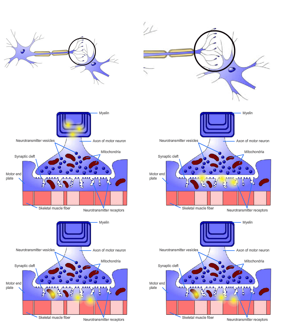
Conduction across a synapse
neurotransmitters are released from the vesicles at the end of the synaptic knob. The most important neurotransmitters are acetylcholine and noradrenaline. This makes the nervous impulse an electrochemical impulse. As vesicles are only found in the synaptic knob at the end of an axon, nervous impulses can only travel in one direction.
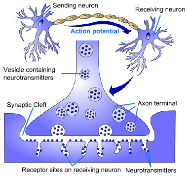
When a signal arrives at a synaptic knob, a neurotransmitter is released into the synaptic cleft and diffuses across the synapse, joining with receptors on the post-synaptic membrane. This causes depolarisation of the membrane and initiates a nerve impulse.
There are 2 types of synapses:
A neuromuscular junction is the synapse between a motor neuron and skeletal muscle. A neuroglandular junction is the synapse between a motor neuron and a glandular cell.
UHI / CC0
(Click image to toggle animation on/off)
- Myelin
- Axon of motor neuron
- Neurotransmitter vesicles
- Mitochondria
- Synaptic cleft
- Motor end plate
- Skeletal muscle fibre
- Neurotransmitter receptors
Excitatory and inhibitory neurotransmitters
Neurotransmitters can have an excitatory or an inhibitory effect and can have different effects at different sites. For example, acetylcholine (ACh) is an excitatory neurotransmitter at neuromuscular junctions.
When a nervous impulse finally reaches its destination – a muscle or gland – a neurotransmitter such as acetylcholine crosses the final synapse to reach the effector.
If the effector is a muscle, this area is known as the neuromuscular junction, where the nervous system and muscular system meet and interact. The motor end plate in the muscle is the area that receives the nervous impulse and this results in an impulse or action potential travelling along a muscle fibre, causing it to contract.
UHI / CC0
Sending neuron > action potential > Receiving neuron
- Vesicle containing neurotransmitters
- Synaptic cleft
- Axon terminal
- Receptor sites on receiving neuron
- Neurotransmitters
Action potentials and polarization
A nerve impulse is an electrochemical disturbance which makes an impulse travel along a nerve from start to finish in one direction only. This disturbance is called an action potential.
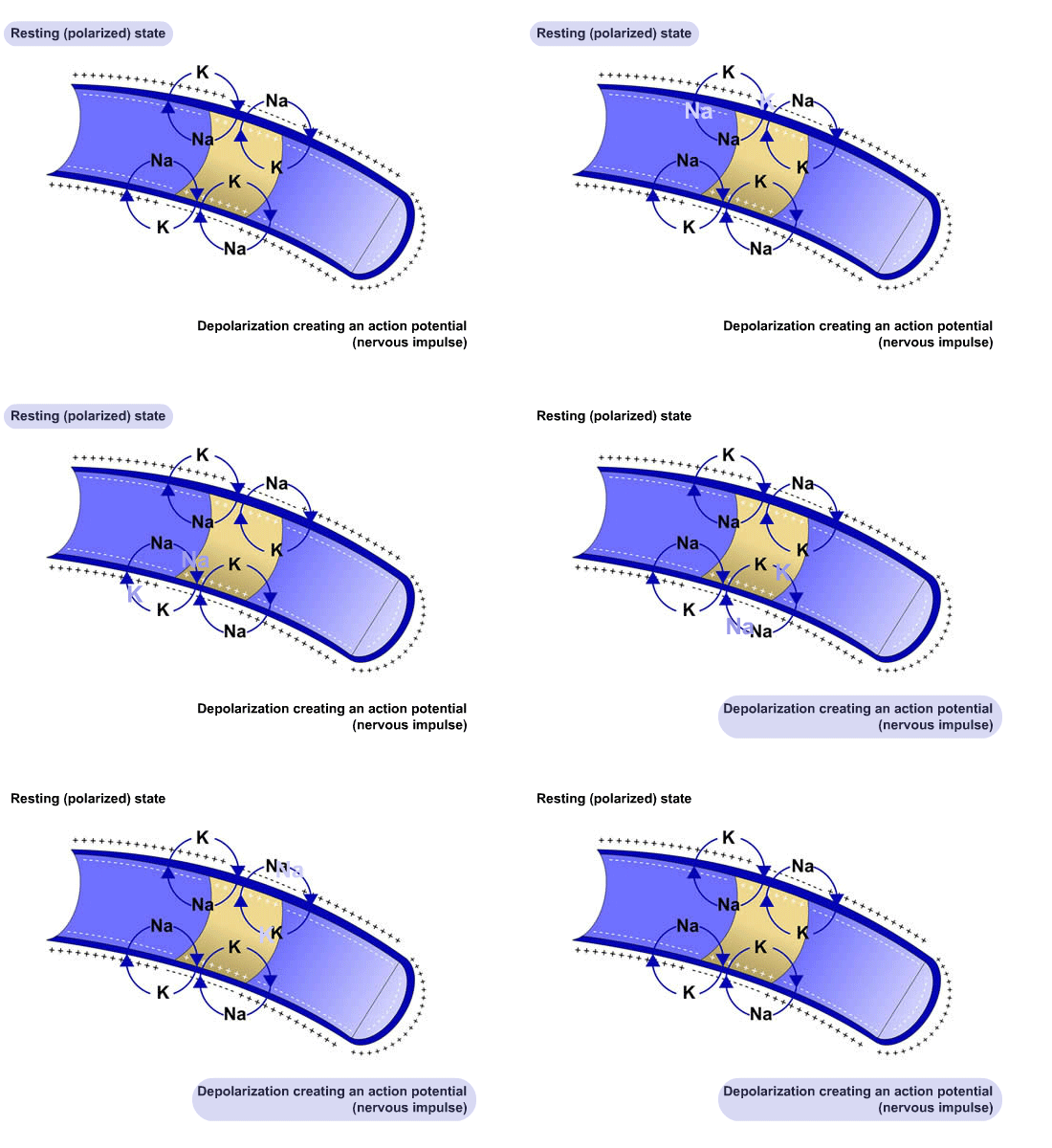
In a resting, un-stimulated state, the axon membrane is selectively permeable - impermeable to sodium ions (Na+) but permeable to potassium ions (K+). This keeps more sodium ions on the outside of the cell membrane and allows entry to more potassium ions. The cell membrane contains more positive charges on the outside and more negative charges on the inside when it is not conducting charges. This is called the resting state or polarised state.
A cellular mechanism known as the sodium pump pushes sodium (Na+) ions out of the cell and draws potassium (K+ ions) in. For every three Na+ ions that are removed from the cell, two K+ ions move in. The resulting imbalance in polarity (electrical state) leaves the inside of the neuron membrane negatively charged compared with the outer membrane surface, as there are more positive ions on the outside of the cell membrane. This potential difference is known as the resting membrane potential (RMP).
UHI / CC0
(Click image to toggle animation on/off)
Resting polarised state > sodium pump pushes sodium (Na+) ions out of the cell and draws potassium (K+ ions) in > depolarisation creating action potential (nervous impulse)
When nerves are excited by a stimulus, this changes the permeability of the neuron, opening up sodium gates that allow the movement of Na+ ions into the cell. This temporarily reverses the polarity across the cell membrane, creating a positive charge on the inside and a negative charge on the outside known as depolarization. This creates an action potential or electrical impulse.
The depolarisation and repolarisation continues along the length of a nerve fibre at each synaptic gap, until it reaches its target muscle or gland.
As the action potential moves along the nerve membrane, the previous point is repolarised and the resting potential returns. At this time the nerve cannot form a new action potential and so this is called the refractory period. If a stimulus is not strong enough to alter the resting membrane potential sufficiently, no action potential is generated.
Quiz
Reflex reactions
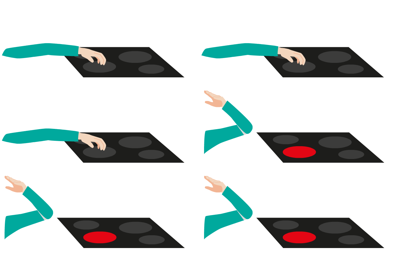
When you touch a hot object you instinctively take your hand away - this is called a reflex action. In a reflex action such as this, the brain is not involved; the impulse travels from a sensory neuron through the spinal cord via a relay neuron to make your muscle move quickly.
Reflex responses often protect the body from damage. They may be coordinated by the brain (cranial reflexes) or the spinal cord (spinal reflexes) but they are involuntary actions that do not involve conscious thought. Reflex responses are the simplest but fastest type of response.
UHI / CC0
(Click image to toggle animation on/off)
Neuronal circuits
The central nervous system contains billions of neurones organised into neuronal pools, each pool with its own sensory, integrative and motor functions.
These pools are arranged in circuits. A simple circuit involves a pre-synaptic neurone stimulating the next neurone and so on, conveying an electrical stimulus (message) from one end of a circuit to the other. However, most circuits are more complex than this.
In converging circuits several pre-synaptic neurones meet a single post-synaptic neurone, in diverging circuits, one pre-synaptic neurone meets several post-synaptic neurones, and in oscillating circuits, a pre-synaptic neurone causes post-synaptic neurones to transmit a series of nerve impulses in a cycle.
UHI / CC0
- Nerve
- Axons
- Blood vessel
- Connective tissue
- Axon terminal
- Schwann cell
- Myelin sheath
- Node of Ranvier
- Cell body
- Axon
- Dendrite
- Nucleus
Converging circuits
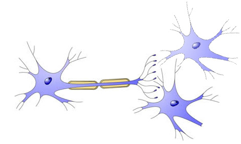
Several pre-synaptic neurones meet a single post-synaptic neurone, allowing more effective stimulation or inhibition of the single neurone receiving the message. The post-synaptic neurone may receive nerve impulses from several different sources.
Diverging circuits
A single pre-synaptic neurone meets several post-synaptic neurones in this type of circuit, allowing a single neurone to stimulate several other neurones, muscle fibres or gland cells simultaneously.
UHI / CC0
Oscillating circuits
In oscillating circuits, once a pre-synaptic neurone is stimulated it causes post-synaptic neurones to transmit a series of nerve impulses. The first post-synaptic neurone is stimulated, and automatically passes the nervous impulse on to a second post-synaptic neurone which does the same, and so on. The stimulation may last from seconds to a few hours, and may be turned off by an inhibitory neurone.
Test your knowledge
Now it’s time to test your knowledge of the nervous system.
There are 10 questions to complete.
You might want to take some time to read your notes, draw diagrams or watch the animations again before you test yourself!
Summary
You have completed your study of the nervous system.
You should now have a good knowledge and understanding of the anatomy and physiology of the nervous system.
You should be able to...
- Outline the organisation of the nervous system
- Identify and name the major parts of a neurone
- Explain the functions of the nervous system
- Explain how the nervous impulse works
 UHI / CC0
