Introduction
You might work through this system over several weeks, days or hours, but to enhance your learning and enjoyment make sure you break it up into bite-size chunks.
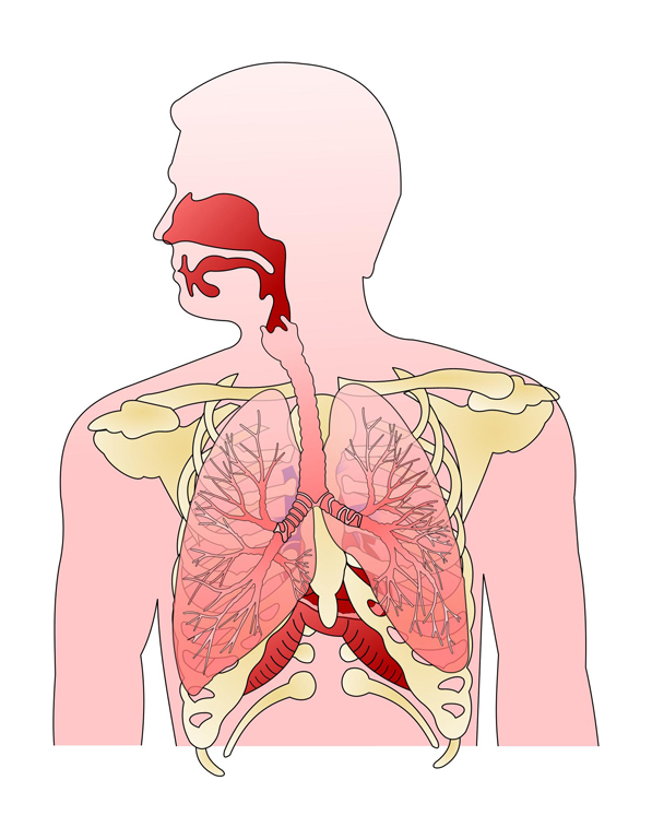
Here are the sections of the respiratory system:
As you study you will learn about:
- Organs of the respiratory system
- Muscles of the respiratory system
- The functions of the respiratory system
- Respiration and ventilation
- Inhalation and exhalation
- Control of breathing
- The pulmonary blood supply
- Gaseous exchange
- Transportation of gases
- Lung capacity terms
 UHI / CC0
Make notes as you study each section, and interact fully with the activities – watch the animations and complete the quizzes.
Take a break at the end of each section – resting your eyes from the computer screen, getting some fresh air or taking a coffee break will improve your ability to focus on your study and take in information.
Give yourself time to think about what you have learned, and time to absorb and understand it.
Anatomy of the respiratory system
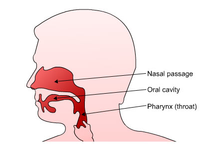
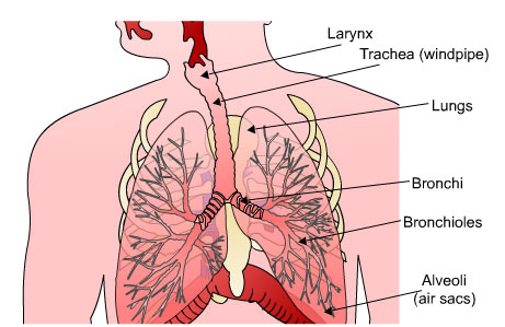
The organs of the respiratory system are divided into the upper and lower respiratory tracts as shown.
Upper respiratory tract
- Nose - nasal passage
- Mouth - oral cavity
- Pharynx (throat)
 UHI / CC0
Lower respiratory tract
- Larynx
- Trachea (windpipe)
- Bronchi and bronchioles
- Lungs
- Alveoli (air sacs)
 UHI / CC0
The upper respiratory tract
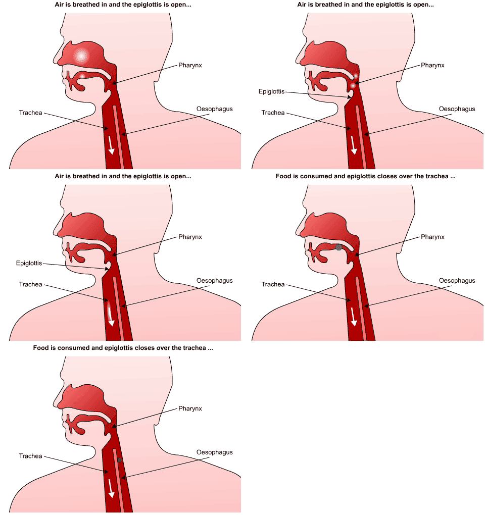
Air is breathed through the nose or mouth, and as it passes through the air passageways in the respiratory system it is warmed to body temperature, moistened and cleaned. Air passes through the pharynx and down the trachea (windpipe). The trachea lies immediately in front of the oesophagus, which is where food and drink is directed into the digestive system. A flap of cartilage called the epiglottis closes over trachea when we swallow, preventing food or drink from entering the lungs.
UHI / CC0
(Click image to toggle animation on/off)
Air is breathed in and the epiglottis is open. Food is consumed and the epiglottis closes over the trachea so food travels down the oesophagus instead.
The nose
At the start of the upper respiratory tract is the nasal cavity which warms, moistens and filters the air we breathe in. The nose is lined with ciliated epithelium which contains mucus secreting cells. The hairs trap the larger particles, and dust and any microbes stick to the mucus. The cilia beat rhythmically which moves the mucus up to the throat where it is swallowed or spat out. The moist mucus also humidifies the inhaled air.
 UHI / CC0
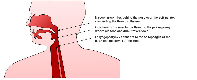
Nasopharynx - lies behind the nose and over the soft palate, connecting the throat to the ear
Oropharynx - connects the throat to the passageway where air, food and drink travel down
Laryngopharynx - connects to the oesophagus at the back and the larynx at the front
The pharynx (throat)
The pharynx is a 13 cm tube consisting of three parts: the nasopharynx, the oropharynx and the laryngopharynx.
The lower respiratory tract
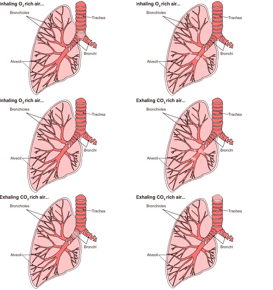
The larynx is also called the voice box, and it connects the laryngopharynx with the trachea. Air passes from the trachea to the bronchi and bronchioles in the lungs. The bronchioles split many times to create a network of vessels throughout the lungs, and at the end of each bronchiole, there are a number of air sacs called alveoli. It is through these air sacs that oxygen diffuses from the lungs via the alveoli into the bloodstream and simultaneously, carbon dioxide diffuses from the blood into the alveoli to be breathed out. The inhaled oxygen is then passed into the blood system where it is transported to the rest of the body.
UHI / CC0
(Click image to toggle animation on/off)
Inhaling O2 rich air: air travels down trachea and bronchi to bronchioles and alveoli.
Exhaling CO2 rich air: air travels from bronchioles and alveoli up through the bronchi and trachea.
The trachea
The trachea or windpipe is composed of cartilage which provides rings of support and prevents it from collapsing. The epiglottis is made of cartilage; it closes off the larynx during swallowing and protects the lungs from inhaling foreign objects such as water or food.
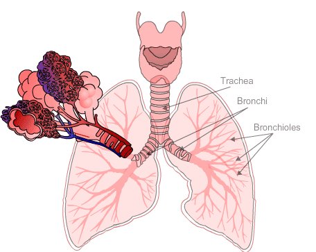
The trachea carries air from the larynx and then divides into two tubes called bronchi. It also contains ciliated epithelium which contains mucus secreting goblet cells. These cells trap microbes and dust and waft it upwards via the cilia to the throat where it can be swallowed.
The bronchi
The trachea divides into two bronchi which contain the same epithelium lining and goblet cells as before.
Each branch of the bronchus subdivides into smaller tubes called bronchioles and finally terminates at the alveolar ducts where the alveoli (air sacs) end the respiratory tract. These are surrounded by capillaries which transfer oxygen and carbon dioxide in and out of the blood.
 UHI / CC0
The Lungs
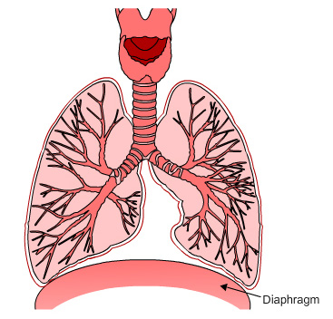
There are two lungs which lie within the ribcage, extending from the diaphragm to just below the clavicle. The base of each lung is concave and fits over the convex area of the diaphragm.
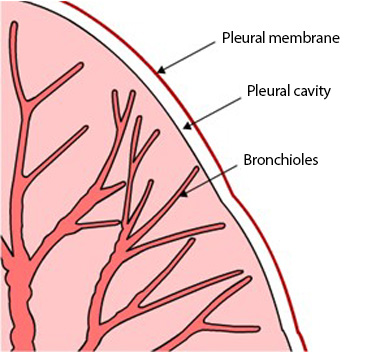
The pleural membrane encloses and protects each lung. Between the membrane and the lung is a fluid-filled space called the pleural cavity which lubricates and reduces friction between the membranes.
 UHI / CC0
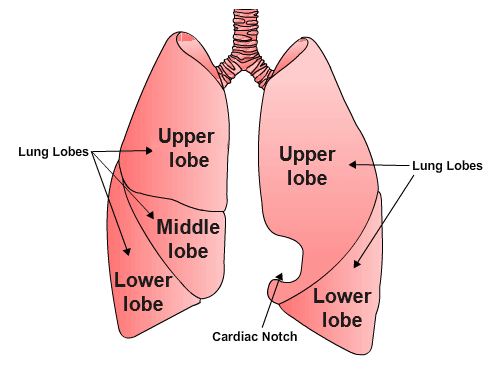
The inner (medial) surface of each lung has an area called the hilus through which bronchi, pulmonary blood vessels, nerves and lymphatic vessels enter and leave the lungs. The lungs are conical in shape and are separated by the heart in the thoracic cavity into two distinct chambers.
The left lung has a concave area called the cardiac notch which creates space for the heart. Each lung has a separate bronchus and is divided into lobes which have many compartments called lobules. Each lobule is served by its own lymphatic vessel, arteriole, venule and a branch from a terminal bronchiole, which then divides into the alveoli.
 UHI / CC0
The alveoli
There are different types of alveoli; whilst most are ideally adapted for gaseous exchange, others secrete alveoli fluid which keeps the alveolar cells moist. Moisture reduces surface tension, making inflation easier and facilitating gaseous exchange.
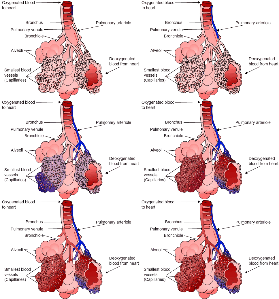
Each lobule arteriole and venule disperses into a dense capillary network which covers the outer wall of each alveoli. With only a single layer of endothelial cells and a basement membrane creating the capillary wall, diffusion of gases from and to the blood is easily facilitated.
Note that the pulmonary arterioles carry deoxygenated blood from the heart, and the pulmonary venules carry oxygenated blood back to the heart – everywhere else in the body arteries carry oxygenated blood and veins carry deoxygenated blood, but the pulmonary circuit between the heart and lungs is different.
UHI / CC0
(Click image to toggle animation on/off)
The pulmonary arterioles carry deoxygenated blood from the heart, inside the alveoli are the smallest blood vessels (capillaries) here the gaseous exchange takes place and then the pulmonary venules carry oxygenated blood back to the heart
Muscles of the respiratory system
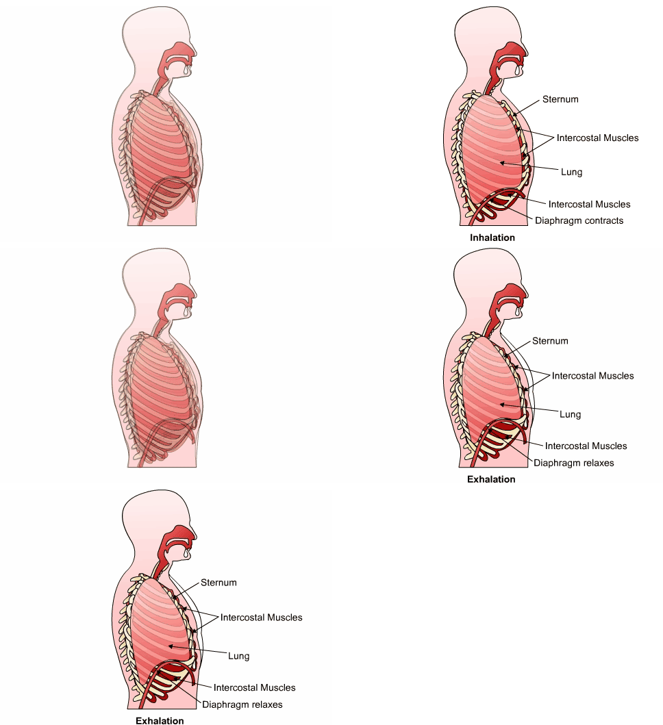
The muscles which are involved in breathing are the internal and external intercostal muscles, and the diaphragm.
UHI / CC0
(Click image to toggle animation on/off)
Sternum, intercostal muscles, lung and diaphragm
Inhalation - Diaphragm contracts
Exhalation - Diaphragm relaxes
Quiz
Now have a go at identifying the organs of the respiratory system. Match the organ names to the corresponding parts.
Functions of the respiratory system
The function of the respiratory system is to provide oxygen for our cells and remove waste products such as carbon dioxide from the body.
We need to breathe in oxygen for all the body’s chemical reactions to take place and to produce energy. Releasing energy from foodstuffs releases carbon dioxide (CO ), and as this is toxic to cells we need to eliminate it as quickly as possible. The respiratory system enables us to take in oxygen via the air we breathe in and expel carbon dioxide as we breathe out.
Respiration and ventilation
Respiration is the exchange of gases between the atmosphere, blood and cells. Inhalation, also known as inspiration, is breathing in;
Exhalation (expiration) is breathing out.
The combination of inhalation and exhalation is also known as ventilation.
Muscles of the respiratory systemThese muscles receive nervous impulse messages from the brain, contracting to initiate inhalation, and relaxing to begin exhalation.
The intercostal muscles are between the ribs - these muscles move the ribcage when we breathe.
The diaphragm is a sheet of muscle beneath the lungs. When the diaphragm is relaxed (during exhalation) it is dome-shaped. During inhalation the diaphragm becomes more flattened.
UHI / CC0
(Click image to toggle animation on/off)
Respiration
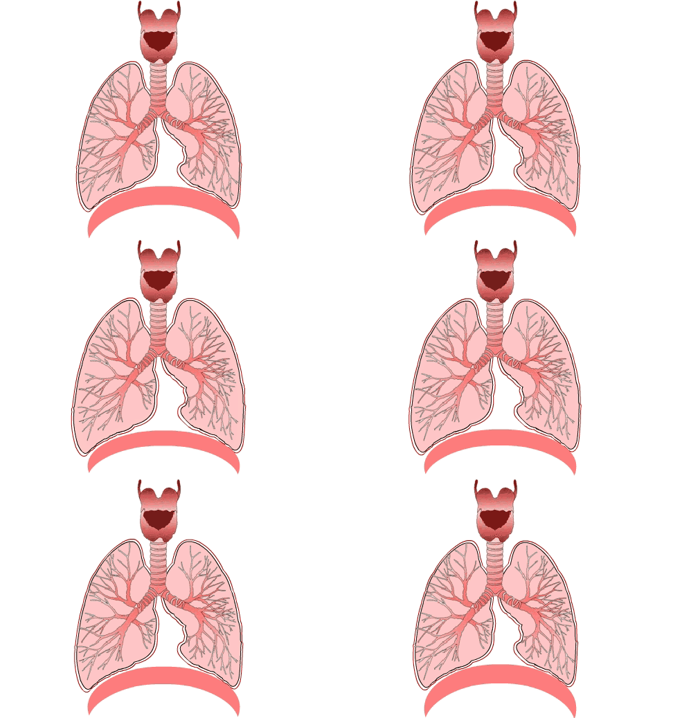
When we breathe, the lungs inflate and deflate aided by muscular activity. The intercostal muscles and diaphragm are the muscles involved in respiration. Although energy is required for the muscular contraction, the process is involuntary.
Air will move into the lungs only when the pressure in the lungs is less than atmospheric pressure when the lungs are deflated. Similarly, air is expelled when the pressure inside the lungs is greater than atmospheric pressure when the lungs are inflated.
UHI / CC0
(Click image to toggle animation on/off)
The respiratory centre
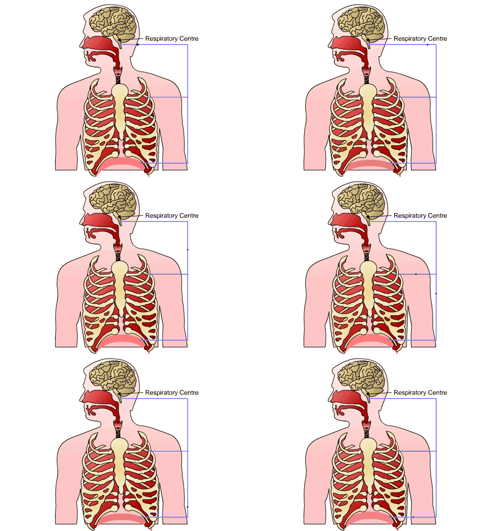
Breathing is an involuntary process that we are not normally aware of, but if we wish to control our rate of breathing, for example, during singing or speaking, then this is a voluntary process (forced breathing). Our breathing is controlled by the brain’s respiratory centre in the medulla oblongata and pons.
The respiratory centre is a group of neurons (nerve cells) organized into 3 areas:
- The medullary rhythmicity area in the medulla oblongata
- The pneumotaxic area in the pons
- The apneustic area in the pons.
The medullary rhythmicity area controls basic rhythmic breathing (inspiration 2 seconds, expiration 3 seconds).
The pneumotaxic area in the pons co-ordinates the transition between inspiration and expiration, ‘switching off’ inspiration before the lungs overfill.
The apneustic area also co-ordinates the transition between inspiration and expiration, activating and prolonging inspiration following nervous stimulation from the pons.

UHI / CC0
(Click image to toggle animation on/off)
Involuntary control of breathing
The basic rhythm of breathing is controlled by autorhythmic neurones in the medullary rhythmicity area. Through a reflex action known as the ‘Hering-Breuer Reflex', nervous impulses are sent to the diaphragm and external intercostal muscles to initiate muscular contraction, and inspiration occurs. At rest, inspiration usually lasts for approximately 2 seconds and expiration for approximately 3 seconds. After 2 seconds of inspiration, the muscles relax and expiration occurs through passive elastic recoil of the lungs and thoracic wall. However, during exercise it is likely that messages from the expiratory area in the brain cause contraction of the internal intercostal muscles and abdominal muscles, causing forced exhalation.
The Hering-Breuer reflex
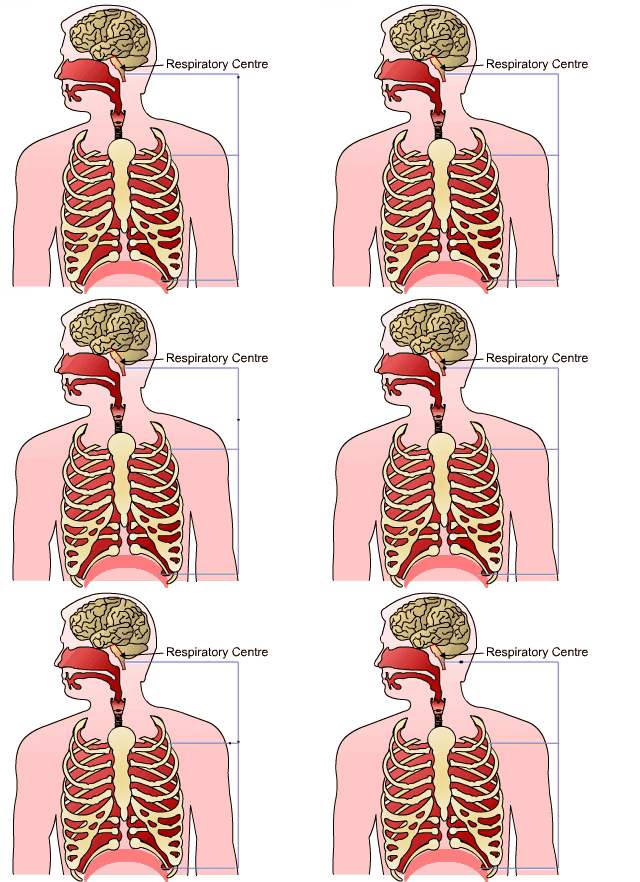
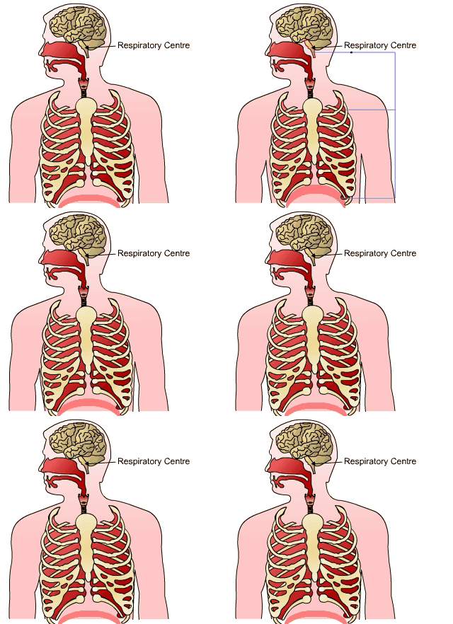
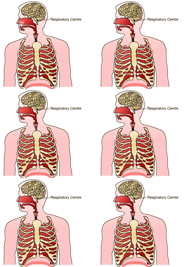
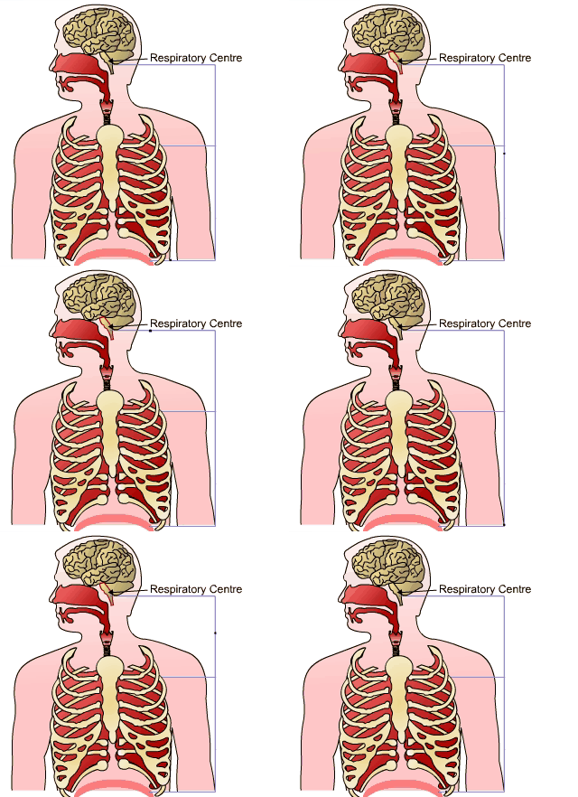
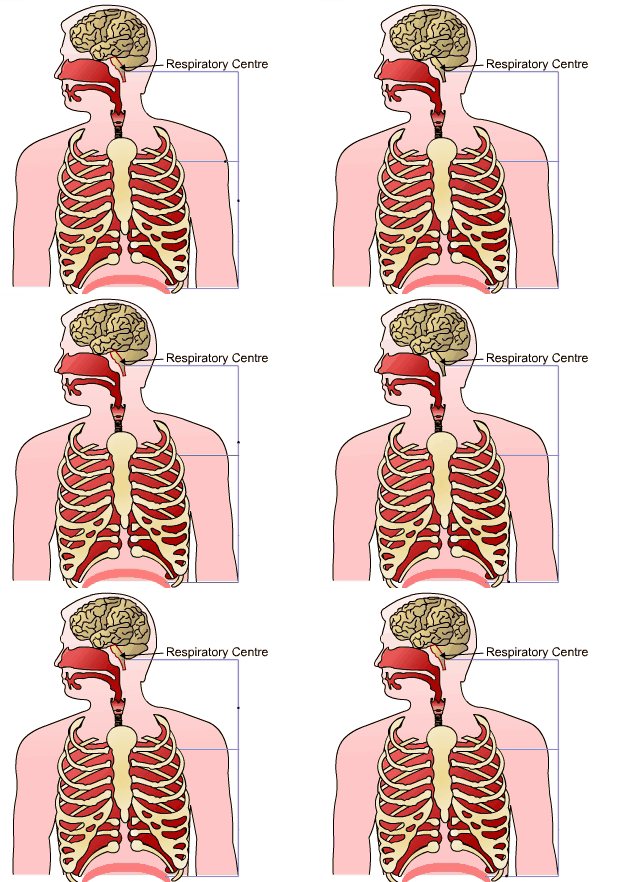
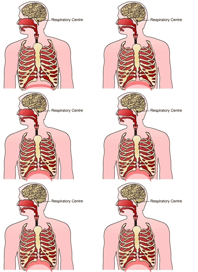
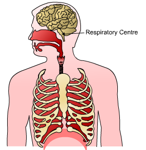
Here are the 7 steps of the Hering-Breuer reflex.
The respiratory centre in the brain sends nerve impulses to the lungs which stimulate the diaphragm and intercostal muscles to contract
 UHI/ CC0
 UHI/ CC0
 UHI/ CC0
As the lungs expand, stretch receptors in the bronchi and bronchioles are stimulates, and send a message to the respiratory centre in the brain.
 UHI/ CC0
 UHI/ CC0
As the intercostal muscles and diaphragm relax, expiration occurs.
 UHI/ CC0
As the stretch receptors are no longer stimulated, inspiration can begin again.
 UHI/ CC0
Respiratory feedback mechanism
Breathing rate can be modified in response to input from other areas of the brain or peripheral nervous system. We can stop breathing if we want to, or control the depth or length of time we inhale or exhale, but this is limited by the build up of carbon dioxide and hydrogen ions in the blood. As levels of these increases, the inspiratory centre in the brain is strongly stimulated to initiate inspiration, and nervous impulses are sent to the inspiratory muscles, overriding our will to hold our breath. Stretch receptors (baroreceptors) in the walls of bronchi and bronchioles also send nerve impulses to inhibit the inspiratory centre in the medulla during over inflation of the lungs, which stops inhalation and prompts exhalation.
Our respiratory system is highly responsive to changes in blood levels of oxygen and carbon dioxide. Chemoreceptors in the medulla oblongata (central nervous system) and in the walls of arteries (peripheral chemoreceptors) relay messages to the respiratory centre in the brain to affect the rate of breathing accordingly.
Oxygen shortage stimulates breathing rate less than increasing carbon dioxide levels, and heart rate usually increases or decreases with respiration rate.
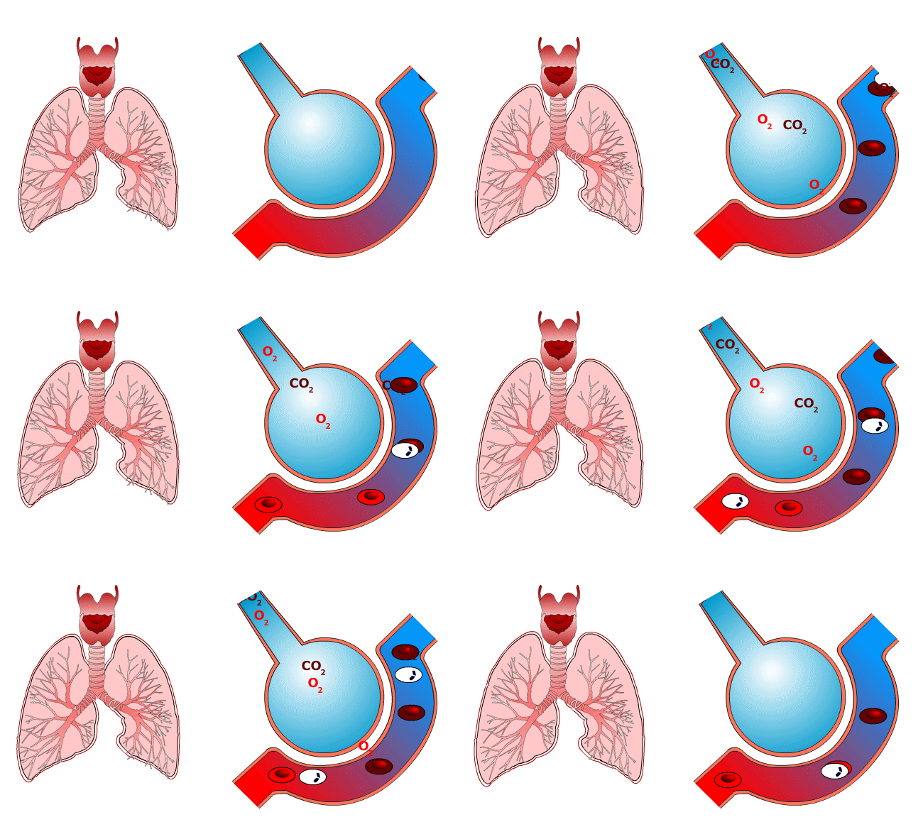
High CO2 / Low oxygen
Rate and depth of breathing increases
UHI / CC0
(Click image to toggle animation on/off)
Low CO2
Rate and depth of breathing decreases
UHI / CC0
(Click image to toggle animation on/off)
Quiz
Pulmonary blood supply
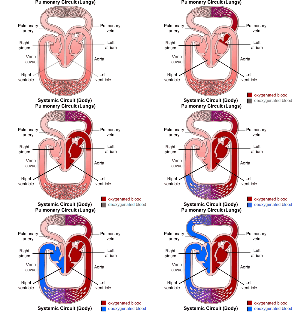
The pulmonary artery from the heart carries deoxygenated blood to each lung.
This artery forms a branched network ending in the capillaries around the alveoli, where gaseous exchange takes place – carbon dioxide diffuses out of the blood and oxygen enters the blood. The capillaries carrying the newly oxygenated blood join up to become the pulmonary vein and send oxygenated blood back to the heart. Note that in the pulmonary cardiovascular system, deoxygenated blood is carried via the pulmonary arteries and oxygenated blood is returned to the heart via pulmonary veins; elsewhere in the body, arteries always carry oxygenated blood and veins carry deoxygenated blood.
UHI / CC0
(Click image to toggle animation on/off)
Pulmonary circuit (lungs)
Oxygenated blood travels from the lungs, through the pulmonary vein, left atrium and left ventricle, through the aorta and into the rest of the body.
Systemic circuit (body)
Deoxygenated blood passes up the vena cavae through the right atrium, right ventricle and pulmonary artery back to the lungs.
Gaseous exchange
The alveoli are particularly well adapted for gaseous exchange:
- They have a large surface area and are moist
- They are thin walled
- They have a good blood supply
- Blood travels slowly through alveolar capillaries maximizing gaseous exchange
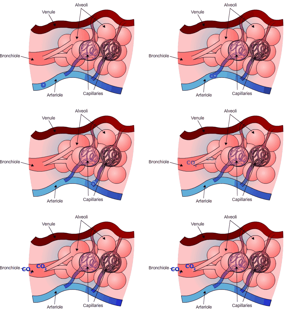
De-oxygenated blood flows from the pulmonary arterioles through the capillaries that cover the alveoli.
By the time the capillary blood is leaving the alveolar surface, much of it is oxygenated and carbon dioxide has diffused from the blood into the alveoli for expiration. The capillaries carrying the newly oxygenated blood join together to form pulmonary venules, carrying the oxygenated blood back to the heart to be pumped around the body.
UHI / CC0
(Click image to toggle animation on/off)
Gaseous exchange and the concentration gradient
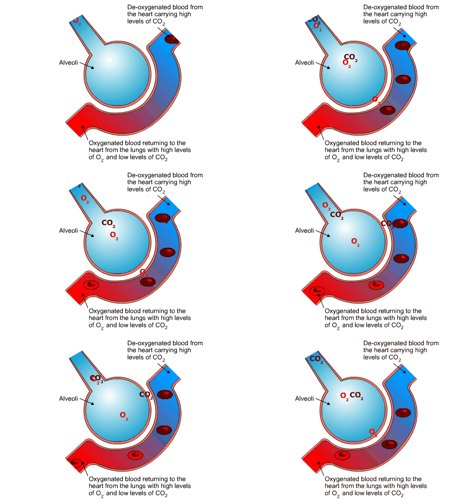
The exchange of gases is by diffusion between the alveoli and the blood capillaries. This is a continuous process and the alveoli are never empty.
As gases diffuse from a high concentration to a low concentration, oxygen diffuses from the alveoli (high concentration of O2) into the de-oxygenated blood (low concentration of O2), and carbon dioxide diffuses in the opposite direction from the blood (high concentration of CO2) into the alveoli (low concentration of CO2).
UHI / CC0
(Click image to toggle animation on/off)
Deoxygenated blood from the heart carrying high levels of CO2 passes the alveoli, CO2 travels from the bloodstream into the alveoli while O2 travels through the thin walls into the bloodstream. Oxygenated blood returns to the heart from the lungs with high levels of O2 and low levels of CO2.
Factors affecting the amount of gaseous exchange
There are a number of factors that affect the amount of oxygen that can be inhaled, and the amount of carbon dioxide that is exhaled.
Surface area of lungs
A person with larger lungs has a greater surface area for gaseous exchange to take place. Males and taller people generally have greater lung size.
Ill Health
Health conditions which affect the respiratory system affect ventilation rate and depth, and/or gaseous exchange. For example, someone with damaged areas of lung tissue due to smoking or disease has a reduced capacity for gaseous exchange.
Fitness Level
The result of training is that oxygen can be delivered to the muscles more efficiently. Recovery after exercise will also be quicker. As we become fitter through continued cardiovascular exercise, our lungs respond to the increasing demands for oxygen, and we also need to be able to expire more carbon dioxide. The number of alveoli increases in the lungs, and we also develop a greater density of capillaries around the alveoli – this enhances the gaseous exchange that needs to take place. Because of these changes, the resting breathing rate is slower, as we are able to take in more oxygen, and expire more carbon dioxide, with each breath.
Partial pressure of gases
If the pressure of oxygen or carbon dioxide increases, the diffusion of that gas also increases. This is noticeable at altitude where there is a lower atmospheric pressure - as the partial pressure of oxygen in the air decreases, this decreases the pressure in the alveoli and reduces the amount of oxygen that diffuses into the alveoli capillaries.
However, over a period of time, the body adapts and increases the number of red blood cells and haemoglobin to accommodate the reduced amount of oxygen available.
When returning to sea level this adaptation remains for a number of weeks and can provide training benefits for endurance sports.
Medication
Some drugs, such as morphine, slow down the rate of ventilation and reduce gaseous exchange.
Quiz
Transporation of gases in the bloodstream
The transport of oxygen and carbon dioxide is vital for respiration to occur. Oxygen is carried around the body as part of a compound called oxyhaemoglobin. This is carried in the blood linked to red blood cells called erythrocytes.
Carbon dioxide is a waste product of metabolism and is carried to the lungs for excretion by three mechanisms:
- In the form of bicarbonate ions in plasma (70%)
- Dissolved in blood plasma (7%)
- By combining with haemoglobin to form carbaminohaemoglobin in red blood cells (23%).
Red blood cells carry heamoglobin. When oxygen has latched onto a cell, the red blood cells (erythrocytes) carry oxyheamoglobin.

UHI / CC0
(Click image to toggle animation on/off)
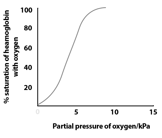
The oxy-haemoglobin dissociation curve
This graph shows that at low oxygen concentrations there is un-oxygenated haemoglobin in the blood and little or no oxyhaemoglobin, but at relatively high oxygen concentrations more haemoglobin in the blood is combined with oxygen, forming oxyhaemoglobin, and able to oxygenate the body tissues.
 UHI / CC0
Lung capacity terms
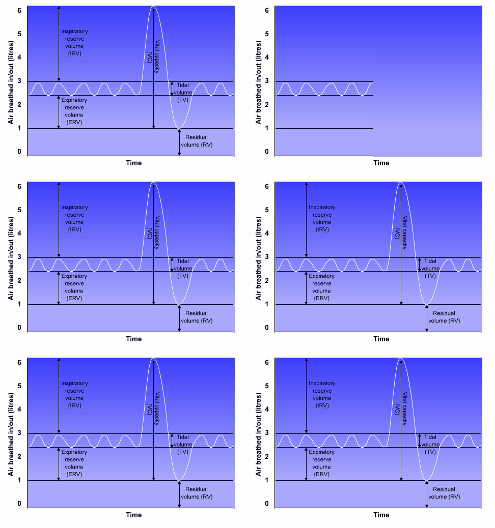
A healthy adult breathes in and out approximately 6 litres of air per minute at rest. Values of lung capacity higher than the norm indicate good pulmonary fitness, and poor values indicate either poor fitness or poor pulmonary function.
Males, younger individuals and those with greater height or body size are likely to have a greater lung volume, though this is taken into account on most ‘norm’ charts.
UHI / CC0
(Click image to toggle animation on/off)
There are a number of lung capacity terms used to describe stages of ventilation.
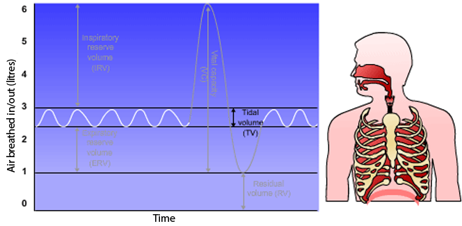
Approximately 500ml of air moves in and out of the lungs with each breath at rest, although this may vary considerably between individuals. This is known as the tidal volume.
 UHI/ CC0
With a deep breath we inhale much more than the amount of air usually inhaled at rest in the tidal volume. The additional amount of inspired air is the inspiratory reserve.
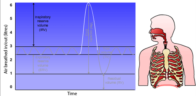 UHI / CC0
We usually exhale approximately 500ml of air with each exhalation, but our expiratory reserve is approximately 1200ml greater. This additional amount is the expiratory reserve volume.
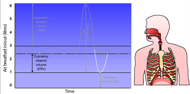 UHI/CC0
This is the total amount of air that can be inhaled and exhaled. Forced vital capacity is usually measured with a spirometer or using a peak flow tube. As well as measuring the volume of air an individual can take in tot heir lungs, as the expiration is forced in this measurement, this also evaluates the strength of the expiratory muscles.
.png?1613473197821) UHI / CC0
UHI / CC0
This is the total amount of forced, expired air in 1 second after maximal inhalation and forced exhalation.
.png?1613473246932) UHI / CC0
UHI / CC0
Even after complete exhalation some air remains in the lung, known as the residual volume.
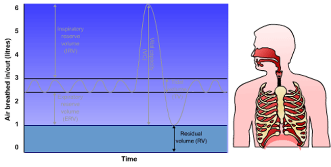 UHI / CC0
This is the sum of the forced vital capacity and residual volume.
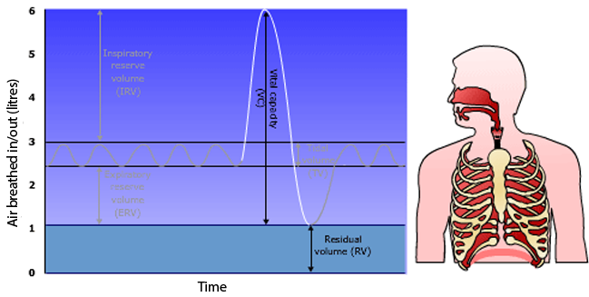 UHI / CC0
Quiz
Test your knowledge
Summary
You have completed your study of the respiratory system.
You should now have a good knowledge and understanding of the anatomy and physiology of the respiratory system. You should be able to:
- Describe the organs and muscles of the respiratory system
- Explain the functions of the respiratory system
- Explain what ventilation, inhalation and expiration are, and how they occur in the body
- Explain the process of gaseous exchange
- Explain how our breathing rate is affected by internal and external factors
- Describe the different lung capacity terms.
 UHI / CC0


 UHI / CC0