Introduction
You might work through this system over several weeks, days or hours, but to enhance your learning and enjoyment make sure you break it up into bite-size chunks.
Here are the sections of the skeletal system:
Bones of the skeleton
As you study the skeletal system you will learn about:
- The skeleton
- Functions of the skeletal system
- Calcium balance
- Classifications of bone
- Bone microstructure
- Ossification
- Joints
- Anatomical terms and terms of movement
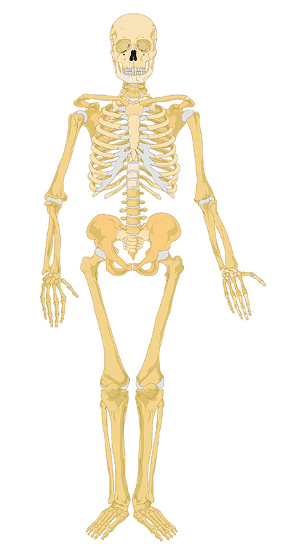
Mikael Häggström/ Public domain
Make notes as you study each section, and interact fully with the activities – watch the animations and complete the quizzes.
Take a break at the end of each section– resting your eyes from the computer screen, getting some fresh air or taking a coffee break will improve your ability to focus on your study and take in information.
Give yourself time to think about what you have learned, and time to absorb and understand it.
Bones of the skeleton
The adult human skeleton is comprised of over 200 bones. Each bone has joints where the bone comes into contact with adjoining bones (articulation).
Bones are attached to neighbouring bones and muscle with connective tissue such as cartilage, tendons and ligaments.
The skeleton is split into bones which form the axial skeleton and bones which form the appendicular skeleton.
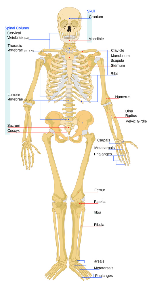
Bones of the axial skeleton form the centre axis of the body and consists of the skull, the vertebral column, the ribs and the breast bone (sternum).
Bones of the appendicular skeleton are bones hanging from the central axis; the shoulder girdle, arms and hands, and the pelvic girdle, legs and feet.
The Skeleton
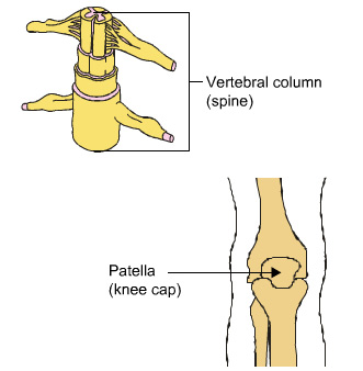
The bones in the axial and appendicular skeleton are listed below, along with their more commonly known names. However, as this is the study of anatomy, you should learn the correct anatomical names for each bone. In addition to the bones listed here, there are also a number of sesamoid bones such as the patella (knee cap).
These are usually nodules of bone embedded in tendons and are thought to modify pressure, reduce friction and alter the direction of the pull of a muscle.
| Axial Skeleton | No. of bones |
|---|---|
| Cranuim (skull) and facial bones | 22 |
| Inner ear bones | 6 |
| Hyoid (found in the throat) | 1 |
| Vertibral column (spine) | 33 |
| Sternum (breat bone) | 1 |
| Ribs (12 pairs on each side) | 24 |
| Appendicular Skeleton | No. of bones |
|---|---|
| Clavicle (collar bone) | 2 |
| Scapula (shoulder blade) | 2 |
| Humerus (upper arm) | 2 |
| Radius and ulna (lower arm) | 4 |
| Carpels (wrist) | 16 |
| Metacarpels (hand) | 10 |
| Phalanges (finger bones) | 28 |
| Pelvic girdles (hip bones) | 2 |
| Femur (upper leg bone) | 2 |
| Patella (knee cap) | 2 |
| Tibia and fibula (lower leg bones) | 4 |
| Talus (ankle bone) | 2 |
| Calcaneus (heel bone) | 2 |
| Smaller heel bones | 10 |
| Metatarsals (foot bones) | 10 |
| Phalanges (toes) | 28 |
The skull
The skull consists of 22 bones and encloses the brain almost completely. It is formed of the following parts:
- the cranium which envelops the brain and bones of the face,
- the mandible (jaw bone).
The top part of the cranium is made up of the frontal bone, the left and right parietal bones and the occipital bone at the back of the skull.
The bones in the cranium are fused together so that there is no movement between them. You will learn more about the different types of joint and the connective tissues that help to create them later on.
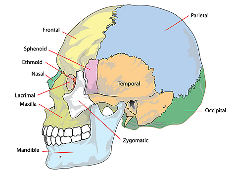
LadyofHats/ Public domain
Parietal
Occipital
Temporal
Zygomatic
Mandible
Maxilla
Lacrimal
Nasal
Ethmoid
Sphenoid
Frontal
Ribs and sternum
The sternum
The sternum (breast bone) is a flat, narrow bone located in the middle of the thoracic cavity.
It is made up of three sections:
- the manubrium at the top,
- the body in the centre and
- the xiphoid process at the bottom.
It articulates with the ribs and the clavicle (collar bone).
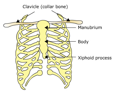
UHI / CC0
The ribs
There are 12 pairs of ribs on each side of the thoracic cavity, increasing in length from the 1st to the 7th rib before they begin to shorten again, giving the characteristic barrel shape.
The upper 7 ribs are connected to the vertebral column at the back and the sternum at the front and are called true ribs.
The remaining 5 pairs of ribs are called false ribs as they are not attached to the sternum directly.
The cartilage of the 8th, 9th and 10th pairs of ribs connect to the cartilage of the 7th pair of ribs.
The lower 2 pairs of ribs do not attach at the front at all and are known as floating ribs.
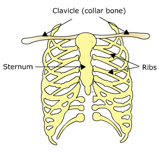
UHI / CC0
The spinal column
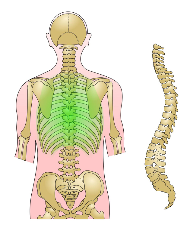
The spinal column is comprised of 33 vertebrae which sit on top of one another creating a supportive column, separated by cartilaginous discs. In the adult the bones of the sacrum and the coccyx at the bottom of the spine fuse together so adults only have 26 vertebral bones.
The functions of the spinal column are:
- To support the upper body, distributing the weight through the pelvis to the legs and feet
- To absorb shock through the intervertebral discs
- To protect the spinal cord
- To allow movement.
The vertebrae
The vertebrae are named depending upon which area of the spine they are in. The cervical vertebrae form the upper end of the spine, the thoracic vertebrae extend through the thorax (chest) region, the lumbar vertebrae form the spine in the small (lumbar arch) of the back, the lumbar vertebrae are positioned between the pelvic bones at the back and are fused together to create the sacrum, and the coccygeal vertebrae form the coccyx at the bottom of the spine.
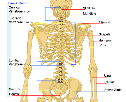
LadyofHats/ Public domain
Spinal column:
- cervical vertebrae
- thoracic vertebrae
- lumbar vertebrae
- lumbar vertebrae
- Sacrum
- Coccyx
- Atlas - top of the neck
Mandible - jaw - Clavicle
- Scapula
- Humerus
- Ribs
- Ulna
- Radius
- Pelvic girdle
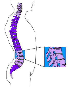
Curves of the spine
As we develop into adults, the spine curves to create the cervical and lumbar curves. These natural spinal curves in the backbone help us function as follows:
- by helping to maintain balance
- by adding strength to the vertebral column
- by absorbing shock as we walk and run
- by helping to protect the vertebral column from fracture.
 UHI / CC0
Quiz
Have a go at identifying each section of vertebrae and see if you can remember how many vertebrae make up each section.
The pectoral girdle
The appendicular skeleton consists of the pectoral and pelvic girdles and the limb bones. The upper (anterior) limbs are attached to the pectoral (shoulder) girdle and the lower (posterior) limbs are attached to the pelvic (hip) girdle.
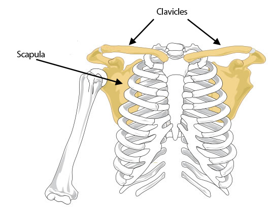
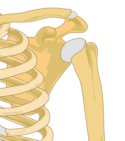
The pectoral girdle consists of two shoulder blades (scapulae) and two collar bones (clavicles). These bones articulate with one another, allowing movement.
LadyofHats/ Public domain
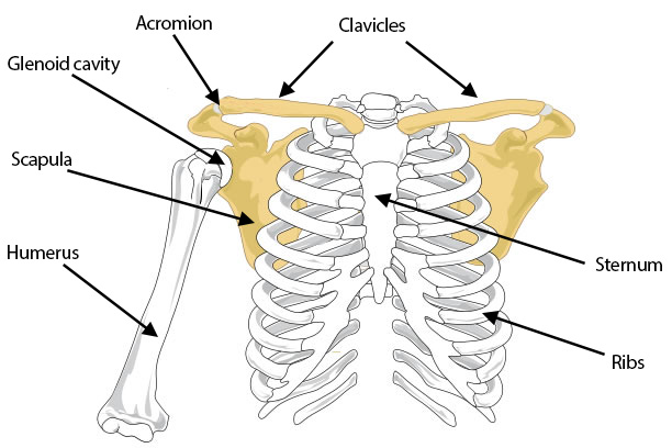
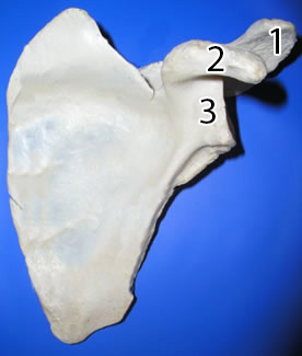
The scapula is a flat triangular bone which stretches from the shoulder to the vertebral column at the back. On the posterior side it has a bony ridge above the shoulder joint called the acromion. This is a place of muscular attachment. Beneath the clavicle on the inside of the shoulder joint, is another bony projection of the shoulder blade called the coracoid process, which also serves for the attachment of muscles. The head of the humerus (upper arm bone) fits into the glenoid fossa of the outer scapula, forming a ball and socket joint.
Key
- Acromial process
- Coracoid process
- Glenoid cavity
Each collar bone is rod-shaped and roughly S-shaped. It lies horizontally and articulates with the upper end of the sternum just above the first rib. The lateral (outer) end of each clavicle articulates with the acromion of the scapula.
The clavicles support the scapulae (shoulder blades), also keeping them back so that the arms can hang freely at the sides of the body. The clavicles form part of the pectoral girdle and keep the shoulder joint in place.
LadyofHats/ Public domain
Labelled diagram showing the clavicles, acromion, glenoid cavity, scapula, humerus, ribs and sternum.
Quiz
Now have a go at identifying the bones of the shoulder girdle and the joints that enable movement.
The arm and hand
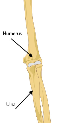
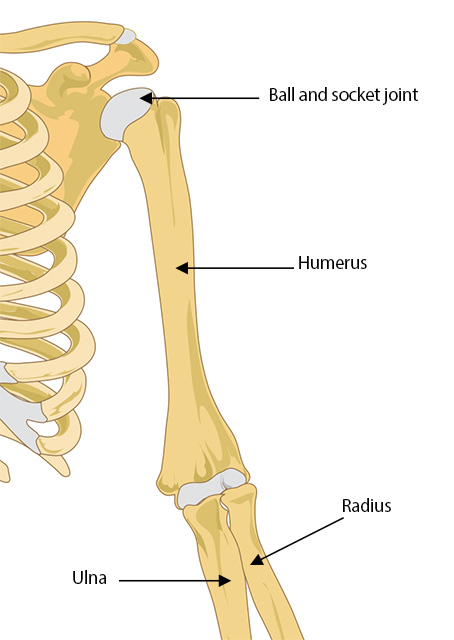
The upper arm is a single long bone called the humerus. The upper end consists of a hemispherical ball which fits into the socket of the shoulder blade to form the shoulder joint. The lower end of the humerus forms a pivot joint with the radius and a hinge joint with the ulna in the elbow.
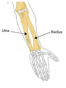
The two long bones of the forearm are the radius and the ulna. The ulna is the larger of the two bones and is situated on the inner side (the little finger side) of the forearm. The upper end of the ulna articulates with the lower end of the humerus forming the hinge joint of the elbow. The lower end of the ulna forms part of the wrist joint.
The radius is situated on the thumb side of the forearm. Its upper end articulates with the humerus and the ulna. The broad, lower end of the radius forms part of the wrist joint, where it articulates with the wrist bones (carpals). The radio-ulnar joints are pivot joints where the moving bone is the radius, allowing the forearm to be rotated.
The Wrist
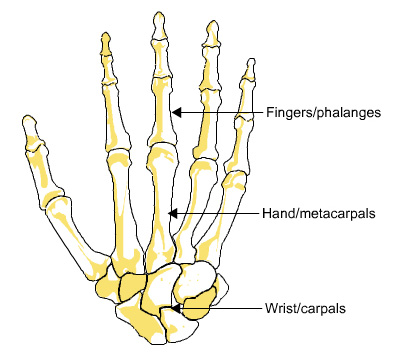
The wrist consists of eight carpal bones. These are small, short bones that are arranged in two rows of four. They slide over one another, creating gliding joints.
The Hand
The palm is supported by five long metacarpals. The metacarpals articulate with carpals at one end and with the phalanges (fingers) at the other end.
The Fingers
The fingers are made up of fourteen phalanges. There are three phalanges in each finger but only two in the thumb.
Quiz
Now have a go at identifying the bones of the arm.
The lower body
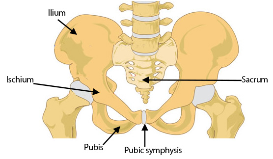
The pelvis consists of two large, sturdy hip bones. Each hip bone consists of three fused bones, the ilium, ischium and the pubis. The ilium is the largest of the three and forms the upper part of the hip bones. The ischium forms the inferior (lower) part of the hip bone, and the pubis is the central front section. The two pubic bones are attached at the front by the pubic symphysis which consists of fibrocartilage and ligaments, and at the posterior via the sacrum forming a complete bony ring. The sacrum has a large, flat surface on each side for articulation with the ilia. On the outer side of the pelvis there is a deep hip socket into which the head of the femur (leg bone) fits.
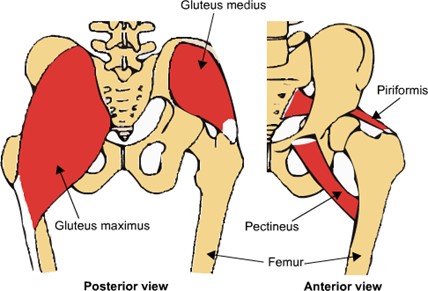
The pelvic girdle forms a strong support for the attachment of the lower limbs. Strong muscles of the back, the legs and the buttocks are attached to it.
UHI / CC0
Posterior view:
- Gluteus medius
- Gluteus maximus
- Femur
Anterior view:
- Piriformus
- Pectineus
- Femur
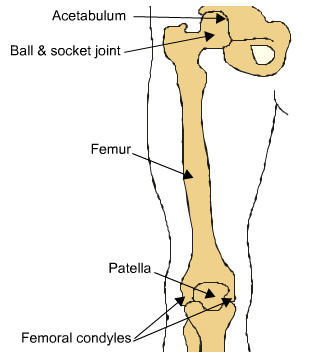
The upper leg bone is the femur and is the longest bone in the body. The head of the femur is turned slightly inwards and has a large, rounded portion which articulates in the acetabulum forming a ball-and-socket joint. At its distal (lower) end, the femur widens to form two large knobs (condyles) which form the hinged knee joint with the tibia (shin bone) of the lower leg.
The patella (kneecap) slides over the anterior (front) of the femoral condyles. The patella is a small, triangular, flat bone which develops on the tendon of the thigh muscle and is attached by ligaments to the tibia.
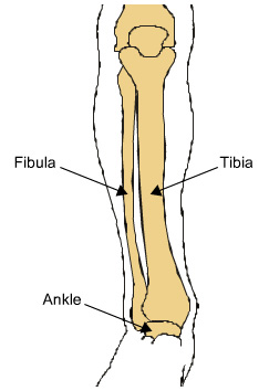
The two bones of the lower leg are the tibia (shinbone) in front and the fibula behind. The tibia is the larger of the two and extends from the knee to the ankle. The upper end of the tibia has two articulating facets into which the condyles of the femur fit to form the knee joint.
The lower end of the tibia articulates with one of the tarsals to form the ankle joint. The fibula is smaller than the tibia and is situated slightly behind it on the outside. The upper end articulates with the tibia but does not form part of the knee joint. The lower end forms part of the ankle joint.
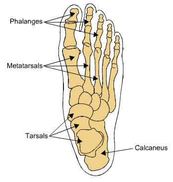
There are seven short, thick tarsal bones, the largest of which is the heel bone (calcaneus) which presses firmly onto the ground when we stand, walk or run. The calf muscles are attached to the calcaneus which allows the heel to be lifted up.
The arch of the foot is formed partly by some of the tarsals but mainly by the five long metatarsals which extend from the tarsals to the toes. The arch is supported by a sheet of connective tissue called the plantar fascia, which helps to support the weight of the body.
Quiz
Now have a go at identifying the bones of the leg.
Skeletal function
Most of the body's tissues are soft and the skeleton provides a rigid but flexible framework and support. Many tissues such as muscles, ligaments and connective tissue are attached to the bones, providing a structure for the body and enabling movement.
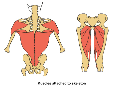
UHI / CC0
The bones, along with the joints and muscles, provide the basis for human movement. The bone provides a lever on which a muscle can pull and, through control from the nervous system, a range of movements, from powerful pole-vaulting to intricate movements, such as speed-typing are carried out.
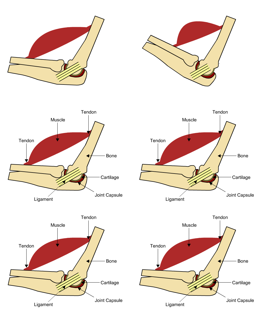
UHI / CC0
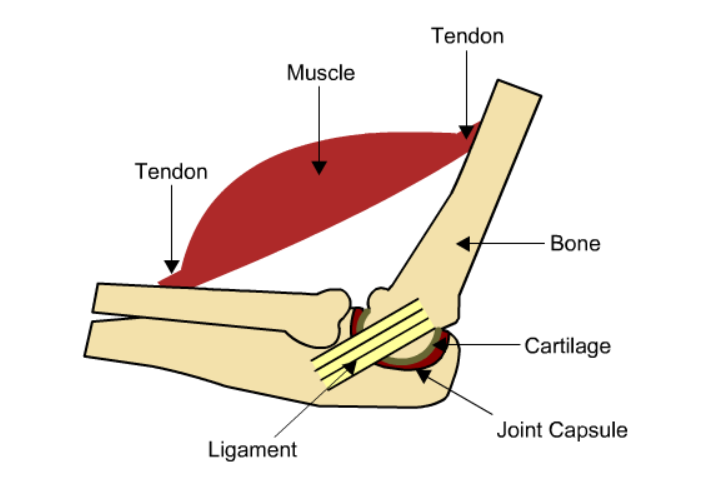
Muscle is attached to bone by tendons, ligament attaches two bones at the joint, joint is protected by cartilage in a joint capsule.
The skeleton creates a number of protective enclosures around the most sensitive of our organs providing a strong protective barrier. The skull almost completely covers the brain, the spinal column protects the full length of the spinal cord, the spine and ribs encase the heart and lungs, and the pelvic girdle helps to protect the bladder and uterus.
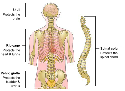
UHI / CC0
- Skull - Protects the brain.
- Rib cage - Protects the heart and lungs.
- Pelvic girdle - Protects the bladder and uterus.
- Spinal column - Protects the spinal cord.
Red blood cells and some white blood cells are formed in bone marrow, which is found within the inside of the bone shaft. Red blood cells are used to transport oxygen around the body and to carry waste products such as carbon dioxide back to the lungs. White blood cells assist as a defence mechanism against infection as part of our immune system.
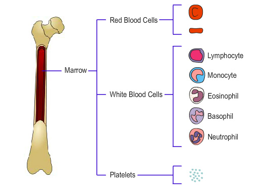 UHI / CC0
Marrow:
- Red blood cells
- White blood cells
- Lymphocyte
- Monocyte
- Eosinophil
- Basophil
- Neutrophil
- Platelets
Mineral storage and calcium balance
Mineral Storage
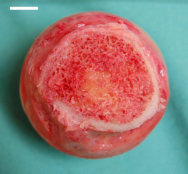
Our bones store minerals such as calcium, phosphate and magnesium and these form part of the bone structure. Bone stores over 99% of the calcium found in the body and blood calcium levels are closely regulated as they heart beat, nerve control, muscle function, blood clotting, blood sugar regulation and many enzymes need it as a co-factor. Calcium is needed for bone and teeth formation, and contributes to the inorganic structure of the skeleton as calcium phosphate, forming a substance called hydroxyapatite.
Calcium Balance
Bone calcium buffers blood calcium levels, providing it when it is needed and re-absorbing it when blood levels are too high. This is known as calcium homeostasis.
The amount of calcium in the bloodstream and the bones is closely controlled by three hormones.
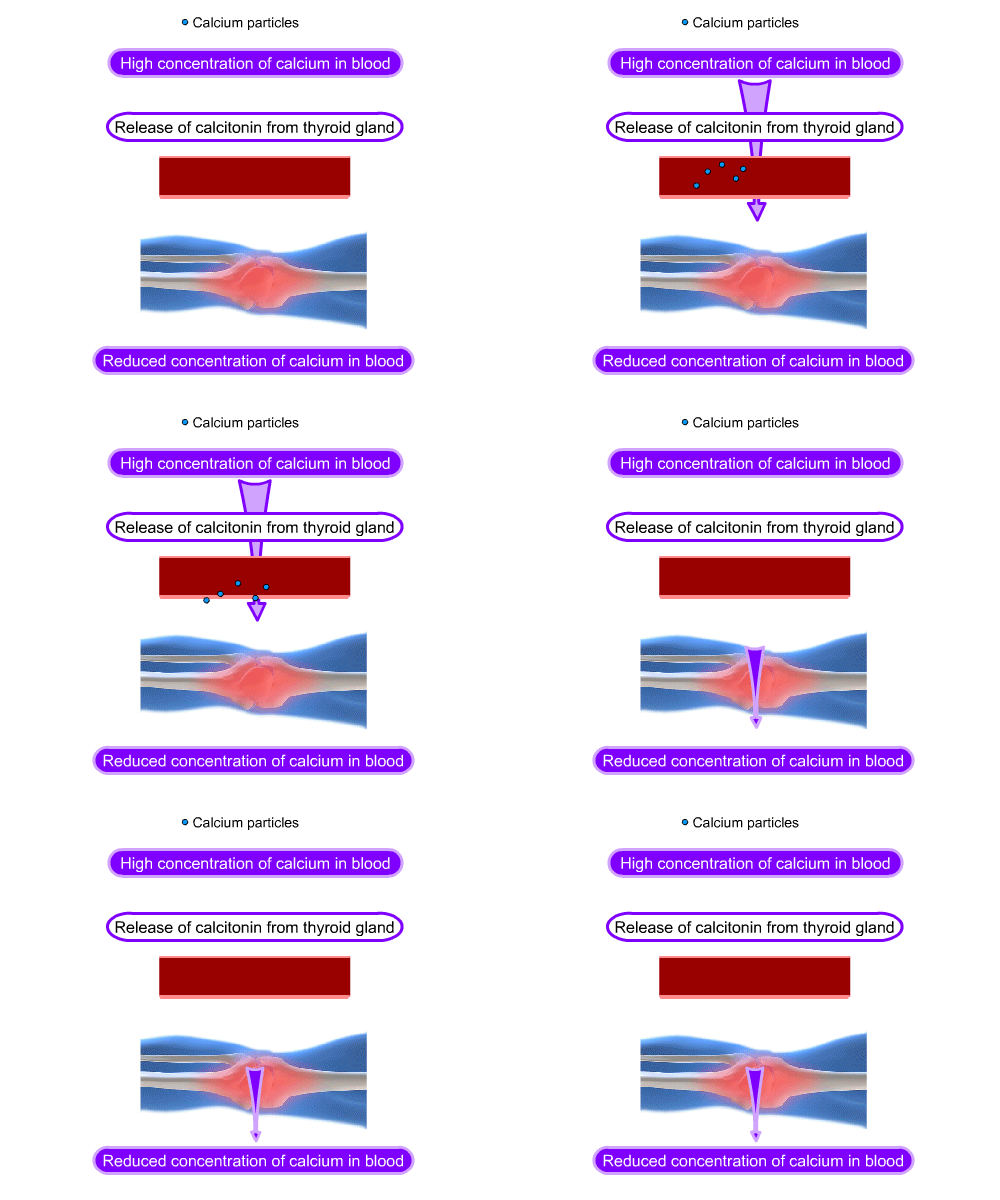
A higher than normal blood calcium level stimulates the parafollicular cells of the thyroid gland to release calcitonin. This hormone increases the amount of calcium deposited in bone, resulting in lower blood calcium levels.
UHI / CC0
Calcium particles
High concentration of calcium in blood
Release of calcitonin from thryoid gland
Reduced concentration of calcium in blood
Calcium homeostasis in the body
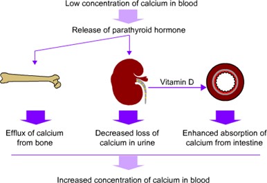
Parathyroid hormone (PTH) is secreted from the parathyroid glands when blood calcium is low. It increases calcium and magnesium resorption from urine in the kidneys, which increases blood calcium. PTH also promotes the formation of calcitriol in the kidneys (synthesised from vitamin D) which increases dietary absorption of calcium in the gut.
If blood calcium levels are still low, parathyroid hormone stimulates osteoclasts (bone cells) to break down bone, resulting in more calcium being released into the bloodstream.
UHI / CC0
Low concentration of calcium in the blood leads to a release of the parathyroid hormone. In bones, this causes an efflux of calcium from the bone. In the bladder, a decreased loss of calcium in urine and production of vitamin D which enhances the absorption of calcium from the intestine. Combined, these processes increase the concentration of calcium in the blood.
Types of skeletal bones
Bones are normally classified according to their shape and size and may be, long bones, short bones, flat bones or irregular bones.
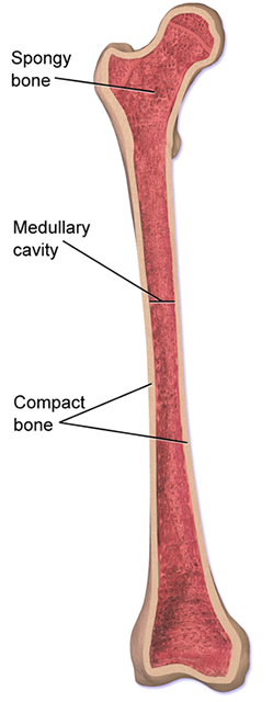
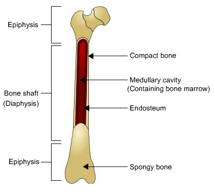
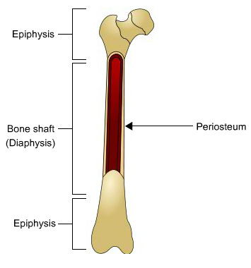
Long bones are longer than they are wide, creating effective levers for movement. Examples of long bones include the humerus, the femur, the tibia and fibula. They consist of a long shaft (diaphysis) with two bulky ends (epiphyses). The epiphyses are the articulating surfaces (surfaces which interact with other joints or bones). Long bones contain some spongy bone, but are primarily made of compact bone, particularly in the middle of the bone shaft where stress is at its highest. Long bones typically have a cavity in the centre of the long shaft called the medullary cavity. This is where bone marrow is stored and it is encapsulated by a layer of connective tissue called an endosteum.
UHI / CC0
Epiphysis
Bone shaft (Diaphysis
Epiphysis
Compact bone
Medullary cavity (containing bone marrow)
Endosteum
Spongy bone
Short bones are usually cube-shaped and are resistant to compression forces. Examples of these bones are the small tarsal bones of the foot (collectively known as the tarsus) and the small bones of the hand and wrist (carpus).
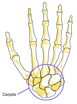
UHI / CC0
These bones often have a protective function. Examples of flat bones are the bones of the skull (cranium), the scapula (shoulder blade), the sternum (breast bone), the ribs and the iliac bones of the pelvis.
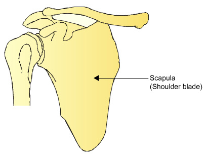
Irregular Bones
Irregular bones are a variety of complex shapes and they may be used for leverage or have a protective or supportive function.
Examples of irregular bones include the vertebral column and the small bones of the face.
Sesamoid bones
Sesamoid bones are bones which are embedded within tendons where pressure is great. The largest bone of this type is the patella (the knee cap).
Quiz
Have a go at identifying the classification of each type of bone.
Microstructure of bone
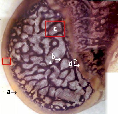
Bone is a solid network of living cells, mineral salts and protein fibres. Bone tissue is made up of bone cells, collagen (connective tissue) and mineral salts.
Bone cells enable bone to function as a living tissue, taking in nutrients from the bloodstream and depositing waste products into the circulatory system to be removed. They also respond to hormones and regulate the amount of calcium that is deposited in, or removed from, the bone matrix.
Mineral salts such as calcium phosphate provide strength and protection. The proteinaceous collagen provides tensile strength and resilience for the skeleton.
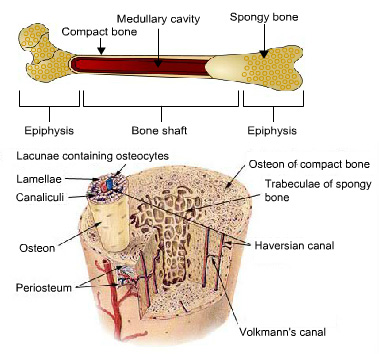 SEER / Public domain
Bone structure
Each bone is surrounded by fibrous connective tissue called the periosteum. This is a tough coating consisting of two dense layers of connective tissue containing a network of blood vessels which supply oxygen and nutrients to the bone, enabling nourishment, growth and repair. The periosteum also provides some protection and enables tendons and ligaments to attach to the bone
Types of bone tissue
Compact bone
Immediately inside the periosteum is a layer of compact bone which forms a hard shell around the outside of the bone, being thickest halfway down the shaft, gradually thinning towards the end of the bone. This provides strength and enables the bone to withstand pressure.
Compact bone tissue forms the outer layer of all bones and is also found in the diaphyses (shaft) of long bones. Compact bone is arranged in rings as shown in this
diagram, and blood vessels, lymphatic vessels and nerves feed bone tissue through penetrating canaliculii.
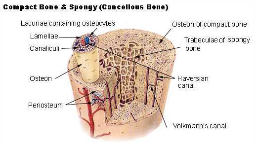 SEER / Public domain
Compact and spongy (cancellous bone)
- Lacunae containing osteocytes
- Lamellae
- Canaliculi
- Osteon
- Periosteum
- Osteon of compact bone
- Trabeculae of spongy bone
- Haversian canal
- Volkmann's canal
Haversian systems
Compact bone is composed of structural units called Haversian systems or osteons. Each osteon unit is made up of cylinders of hard, calcified matrix (hydroxyapatite) called lamellae. In the middle of each cylinder is a narrow channel called a Haversian canal that contains blood vessels and nerves. This allows blood to be supplied to the bone tissue, providing nutrients and minerals to enable bone cell function and bone
mineralization. Blood vessels run through interconnected Haversian canals creating a network that nourishes the bone. Lacunae are small spaces between the calcified rings containing osteocytes
Cancellous (spongy) bone
Inside the layer of compact bone is cancellous or spongy bone tissue, which has the same structure as compact bone but has more spaces between bone tissue filled with blood vessels, fat, fluid and connective tissue. Spongy bone is found mostly in the centre of bones or at the epiphyses (ends of long bones) underneath a thin layer of compact bone and is well suited to cope with the compression forces produced by skeletal weight-bearing.
Key
- Compact bone
- Cancellous bone
- Red marrow cavity
- Epiphiseal plate
*Image used under copyright exception [teaching].
Quiz
Before you go on, have a go at labelling this diagram which illustrates bone structure by dragging the terms to the correct areas highlighted.
Types of bone cell
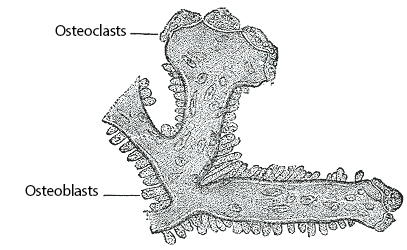
Osteocytes are mature bone cells which maintain bone tissue through the exchange of nutrients and waste. They are derived from osteoblasts and are embedded in compact and spongy bone.
Osteoblasts are bone cells that lay down bone minerals. They make collagen and hydroxyapatite (mineral matrix).
Osteoclasts are bone cells that resorb bone minerals, reducing the amount of calcium and other minerals and salts in bone tissue. They also make collagenase and secrete acids which dissolve hydroxyapatite structure.
Osteoblasts and osteoclasts balance bone tissue being built up or broken down, controlled by calcitonin, parathyroid hormone and calcitriol.
 Henry Vandyke Carter / Public domain
The medullary cavity and bone marrow
Inside spongy bone and in the middle of long bone shafts (diaphyses) is the medullary cavity which contains bone marrow. It is also called the marrow cavity. There are 2 types of bone marrow in most bones.
Yellow bone marrow is found in most bones in the medullary cavity, usually filling the shafts of long bones. It is made up of blood vessels, nerve cells and fat cells (adipose tissue) and can be converted to red bone marrow and produce blood cells if severe blood loss occurs.
Red bone marrow is found mostly in spongy bones and the end of long bones. It produces red blood cells (erythrocytes), white blood cells (lymphocytes) and platelets.
Quiz
Before you go on, have a go at completing this paragraph to test your knowledge of bone structure. If you need a hint, click Reveal to see the missing word options.
compact
Compact
spongy
oesteon
Haversian Systems
spongy
endosteum
osteons
lacunae
lamellae
bone marrow
compact
Haversian
periosteum
medullary cavity
epiphysis
Ossification
Ossification is the name given to bone formation. Depending upon where and when new bone tissue is formed, different names are used to describe the ossification.
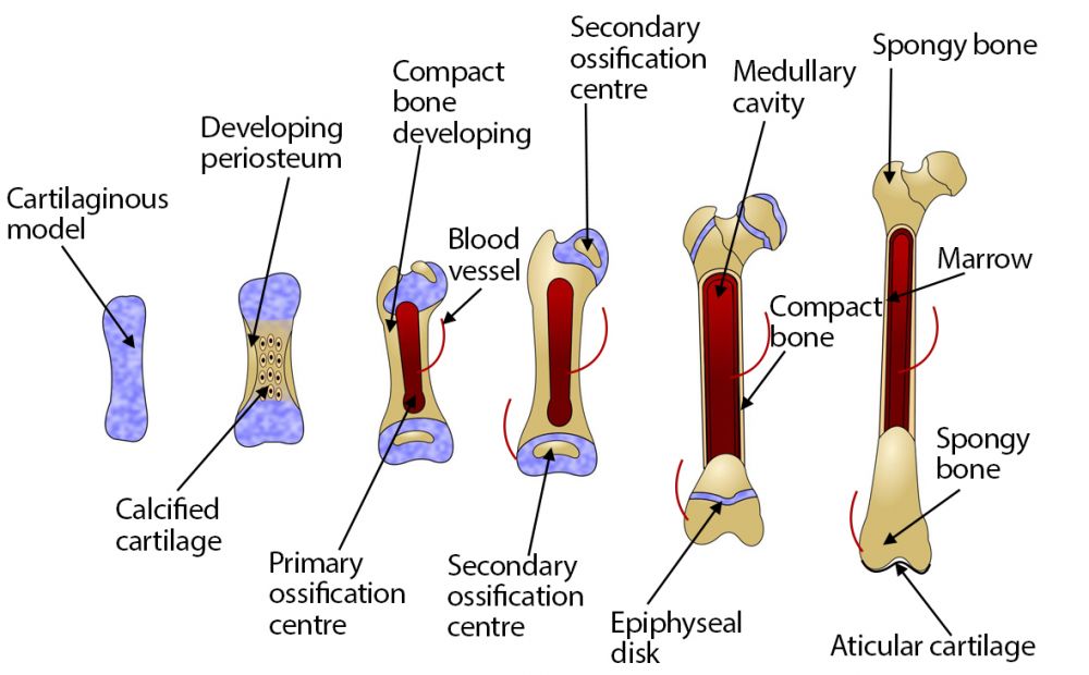
The skeleton of a human embryo is composed of fibrous connective tissue and hyaline cartilage that is shaped like bones – these provide the skeletal structure for ossification to take place. Ossification begins approximately six or seven weeks into embryonic development and continues throughout life; the bones of an infant only become completely hardened by bone tissue during late adolescence. There are two forms of ossification.
Intramembranous ossification refers to bone formed directly onto loose connective tissue without going through a cartilaginous stage (skull, mandible & clavicles).
Endochondral ossification refers to bone formation in hyaline cartilage – this is how most bones, particularly long bones, are formed.
 UHI / CC0
Stage 1 = Cartilaginous model
Stage 2 = Developing periosteum and calcified cartilage
Stage 3 = Compact bone developing, blood vessel and primary ossification centre
Stage 4 = Secondary ossification centre developes
Stage 5 = Epiphyseal disc, medullary cavity and compact bone develop
Stage 6 = Spongy bone, marrow and aticular cartilage
Although these are different methods of bone formation the end result is the same, with either method replacing connective tissue with bone. Most bone formation is endochondral ossification. This type of ossification is split into two phases – Primary and Secondary ossification.
Primary ossification
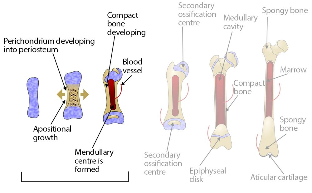
Primary ossification occurs before the epiphyses of bone and the epiphyseal growth plates are formed. A cartilage matrix is initially formed in the shape of bone and surrounded by a membrane called the perichondrium. The cartilage matrix grows and more cells are formed. Most of the growth is an increase in length, known as interstitial growth, but an increase in thickness is called appositional growth. As the cartilage cells grow, the matrix pH changes and calcification begins.
 UHI / CC0
At the same time, blood supply penetrating through the perichondrium stimulates osteoblast formation, and cartilage cells are gradually replaced by osteocytes.
The osteoblasts create a calcified bone matrix known as hydroxyapatite, and the spongy bone originally formed in the centre of the bone is broken down by osteoclasts to form the medullary cavity, which fills with red bone marrow. Primary ossification proceeds inwards from the periosteum.
Secondary ossification
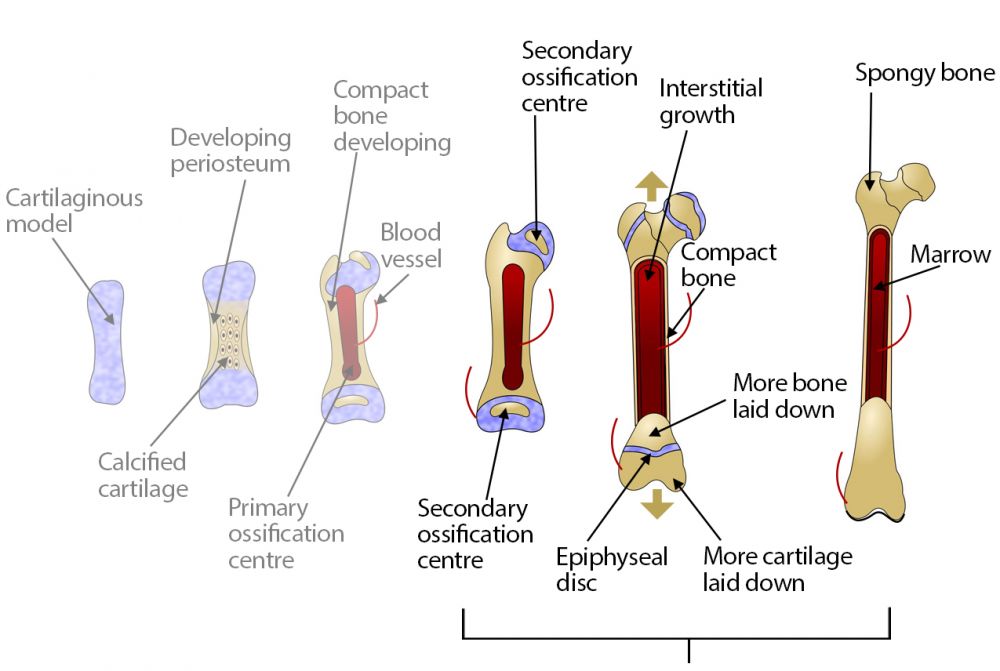
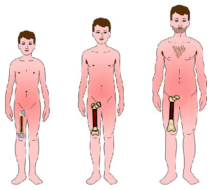
The epiphyseal growth plate or disc (ossification centre) develops shortly after birth where the shaft (diaphysis) and the epiphyses meet in long bones, and this is where interstitial bone growth occurs.
Long bones develop and grow throughout childhood at these growth plates at each end of the bone. Bone grows in length as more cartilage (matrix & chondrocytes) is produced on the epiphyseal side and bone is laid down on the diaphyseal side of the growth plate.
 UHI / CC0
The growth plates consist of cartilage in children and young adults, but as we mature this is replaced by bone. If the epiphyseal plate is damaged during childhood by fracture or as a result of repetitive or demanding exercise, the bone may be shorter, as any damage to the cartilaginous growth plate results in the growth plate closing earlier.
Ossification continues until the early twenties when all epiphyseal disc cartilage is replaced by bone (closure of the epiphyseal disc) and the bone lengthening process stops.Completion of ossification
Bones begin to fuse prior to teenage years but ossification is not complete until an adult enters their twenties. Growth usually stops at approximately age 18 in females and age 21 in males. Although ossification ends, bone is a living tissue, and as such, it is constantly being broken down and rebuilt.
In adults, cartilage is still found in the tip of the nose, the external ear, the voice box (larynx), and in joints (knee). Cartilage is also found where the ribs are attached to the breastbone (sternum), thus allowing the rib cage to move during breathing.
 UHI / CC0
Quiz
Before you go on, have a go at this quiz to test your knowledge on ossification by putting the words to the correct areas. If you need a hint, click Reveal to see the missing word options.
epiphyseal growth plate
appositional growth
medullary cavity
epiphyseal
skull
Primary ossification
cartilage
bone
endochondral ossification
perichondrium
diaphyseal
interstitial growth
Intramembranous ossification
cartilage
Secondary ossification
Anatomical terms
The following terms are commonly used when referring to any part of the body, or to determine body movements. Familiarize yourself with these words, as you will come across these terms as you study the joints and the muscles of the body.
Anterior: To the front
Posterior: To the back
Lateral: To the side
Distal: Furthest away from
Medial: Closest to
Superior: Above
Inferior: Below
|
Movement |
What happens |
Example |
|---|---|---|
| Flexion | The angle at a joint reduces | Biceps muscle bending elbow |
| Extension | The angle at a joint increases | Triceps muscle straightening the elbow |
| Abduction | Body parts move away from one another or away from the body | Lifting your arm or leg outwards away from the body |
| Adduction | Body parts move closer together or towards the middle of the body | Bringing the arm/leg back in |
| Circumduction | A body part rotates forming a cone shape or large circle | Circling an outstretched arm in a circle from the shoulder (windmill action) |
| Medial rotation | A bone moves around its long axis towards the body | Rotating the thigh or arm inwards. Keeping arm by side, twist inwards so the palm of the hand faces backwards |
| Lateral rotation | A bone moves around its long axis away from the body | Rotating the thigh or arm outwards. Keeping arm by side, twist outwards so the palm of the hand faces the front |
| Dorsiflexion | Pointing foot/toes upwards | Flexing foot |
| Plantar flexion | Pointing foot/toes downwards | Pointing toes |
| Elevation | Lifting upwards (shoulders) | Shrug shoulders up |
| Depression | Movement of body part down | Shoulders back down |
| Protraction | Moving shoulder girdle forwards | Turn shoulders inwards towards centre of chest |
| Retraction | Moving shoulder girdle backwards | Move shoulders back (e.g. when sitting or standing up straight) |
| Lateral flexion | Bending sideways around the vertebral column | Side bend from standing position (moving upper body over to side from waist) |
| Pronation | Turning the palm of the hand downwards | Turn hand as if you are putting your hand out to receive something |
| Supination | Turning the palm of the hand upwards | Turn hand over so the palm is down |
| Inversion | Turning the soles of the foot inwards | Turn foot inwards as if looking at the sole of the foot |
| Eversion | Turning the soles of the foot outwards | Turn foot outwards as if looking at the sole of the foot |
Joints
Joints occur whenever two or more bones meet at the same point. Bone surfaces at a joint are covered with articular cartilage which provides a smooth surface for movement.
Joints are classified based upon whether there is a space between the articulating bones and the type of connective tissue connecting the joint.
Joints such as the sacrum or lower lumbar vertebrae are fused joints, which offer no movement.
 UHI / CC0
Ball and socket joint
Pivot joint
Saddle joint
Elipsoid joint
Hinge joint
Plane joint
There are three classifications of joints in the human body.
Fibrous (fixed) joints are held together by collagenous connective tissue and there is no cavity, as shown in the cranium.
Cartilaginous joints are slightly moveable – these have no cavity and bones are connected together by cartilage as in the symphysis pubis in the pelvic girdle.
The third and most common classification of a joint in the human body is the synovial joint.
Synovial joints
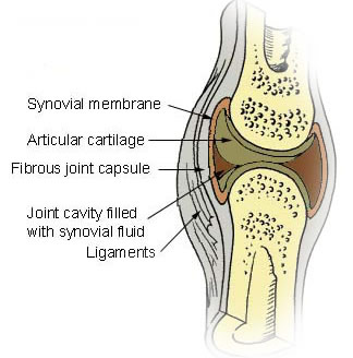
Synovial joints are the most common type of joint found in the musclo-skeletal system. They have a synovial cavity, and the bones are usually surrounded by an articular capsule and ligaments.
Where the bones of a freely movable joint rub together, they are covered with shiny, slippery cartilage called hyaline cartilage and the joints are lubricated by a liquid called synovial fluid. This fluid is sealed in by the synovial membrane which surrounds the whole joint. The bones of freely movable joints are held in place but allowed to move freely by bands of connective tissue called ligaments which join one bone to another.
Types of synovial joint
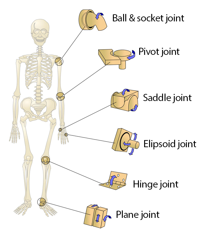
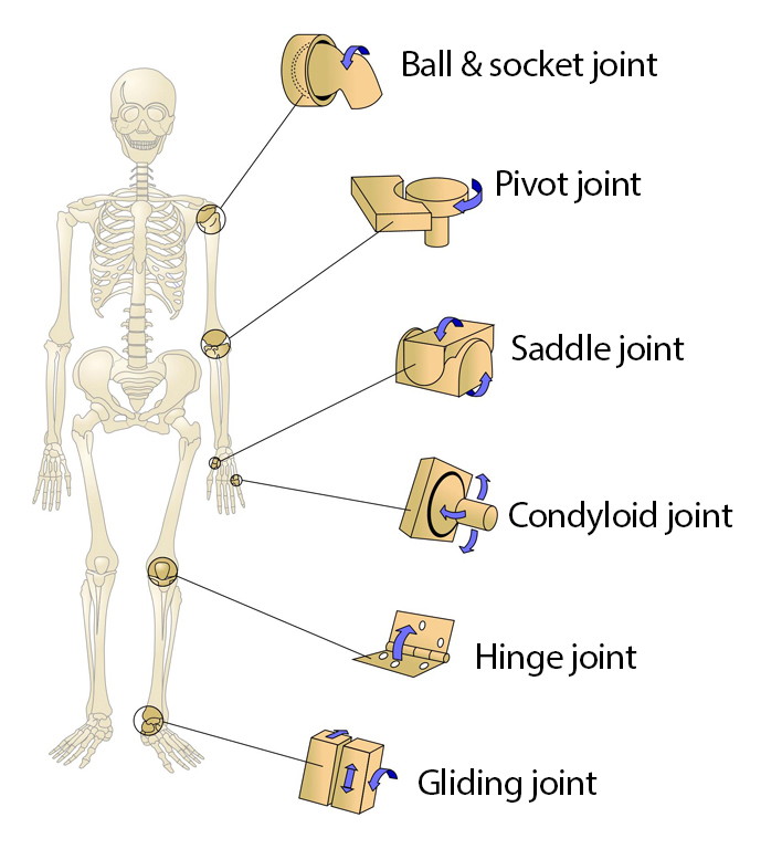
Synovial joints are classified according to their structure and the movement they allow. Here are the 6 types of synovial joints
Ball and socket joint
Pivot joint
Saddle joint
Condyloid joint
Hinge joint
Gliding joint
Tendons and ligaments
Tendons and ligaments are both formed from connective tissue. Tendons join muscles to bone, and ligaments join bone to bone, so they are usually found where muscles cross a joint and where two bones meet. For example, the tendon connecting the quadriceps muscles to the tibia goes through the knee joint, and collateral and cruciate ligaments are also present within the structure of the knee joint providing additional strength and protection for the joint.
 UHI / CC0
UHI / CC0
Tendon
Muscle
Tendon
Bone
Carilage
Joint Capsule
Ligament
Tendons are formed from the outer layers of connective tissue surrounding muscle fibres and connect the end of the muscle to the bone to allow movement when the muscle contracts.
Ligaments connect one bone with another and also increase joint stability. The fibres of the outer layer of a ligament grow into the periosteum of the bone itself, forming a very strong bond. The attachments of ligaments and tendons can be so strong that a muscular contraction or ligament sprain can take a piece of bone away rather than rupture the fusion of the connective tissue with the periosteum.
Quiz
Now test your knowledge of synovial joints.
Factors affecting joint mobility
There are several factors which affect the amount of mobility and flexibility at a joint.
- Shape and compatibility of the joint surfaces
- Impingement of bony structures around a joint
- The limiting effect of muscles, skin and adipose tissue
- Temperature
- Increasing age
Mikael Häggström / Public domain
UHI / CC0
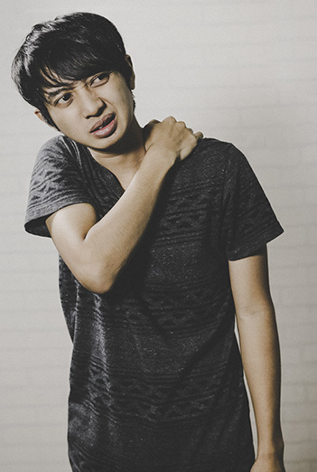
Inflexible, tight muscles may limit the movement at a joint. For example, putting on socks may be difficult with tight hamstring muscles at the back of the leg. This may also be difficult with excess adipose tissue (fat) around the middle. Muscular bulk can occasionally limit full-range movement if muscles are either inflexible of hypertrophied (very large).
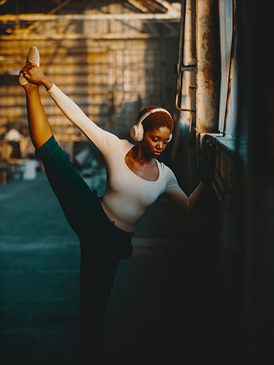
The joint capsule and ligaments all prevent undue joint movement which might lead to damage. Ligaments are utilized more during fast, unplanned movements such as twisting an ankle or in positions where the joint is placed under pressure for example in extreme yoga positions demanding good joint flexibility.
Research indicates that it is easier to mobilize a joint when it is warm. At temperatures above 40°C it is possible to obtain a 20% increase in mobility compared with a normal room temperature but as temperatures fall this mobility is progressively reduced. Warming up prior to exercise helps mobilise joints and increases flexibility - a warmer environment can enhance the flexibility required for some activities.
 Avi Richards / Unsplash
Test your knowledge
Now it's time to test your knowledge of the skeleton system. There are 10 questions to complete.
You might want to take some time to read your notes, draw diagrams or watch the animations again before you test yourself!
Summary
You have completed your study of the skeletal system.
You should now have a good knowledge and understanding of the anatomy and physiology of the skeletal system. You should be able to...
- Name the bones of the skeletal system
- Explain the functions of the skeletal system
- Explain calcium balance
- Describe the classifications of bone
- Describe bone microstructure
- Describe ossification
- Describe the different joints
- Explain anatomical terms and terms of movement
If you think that your knowledge or understanding of any section of the skeletal system could be improved further, go back to the relevant sections and work through them again, taking time to make notes and complete the activities.
 Mikael Häggström/ Public domain


 UHI / CC0