Introduction
You might work through this system over several weeks, days or hours, but to enhance your learning and enjoyment make sure you break it up into bite-size chunks.
Here are the sections of the muscular system…
Make notes as you study each section, and interact fully with the activities – watch the animations and complete the quizzes.
Take a break at the end of each section– resting your eyes from the computer screen, getting some fresh air or taking a coffee break will improve your ability to focus on your study and take in information.
Give yourself time to think about what you have learned, and time to absorb and understand it.
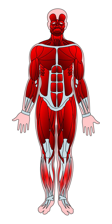
UHI / CC0
Types and characteristics of muscle tissue
Types of muscle tissue
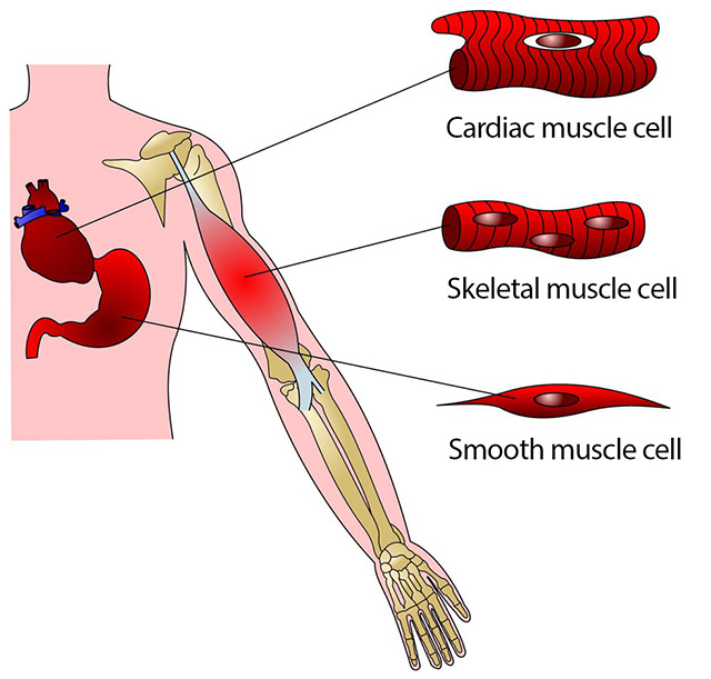
There are three main types of muscle tissue found in the human body:
- cardiac tissue
- smooth muscle tissue
- skeletal muscle tissue
Cardiac muscle is found in the heart, and although it has some similarities to skeletal muscle, it is a specialised type of muscle tissue.
Smooth muscle is found in the walls of arteries and veins, in the bronchioles (in the lungs), and in the digestive tract. Both of these types of muscle tissue are under involuntary control of the autonomic nervous system, whereas skeletal muscle is under voluntary control.
Here, we are studying skeletal muscle.
Characteristics of muscle tissue
All types of muscle tissue share the following characteristics:
Excitability
Muscle tissue responds to stimuli from hormones or neurotransmitters and produces electrical signals called action potentials.
Conductivity
This is the ability of a cell to conduct action potentials along its plasma membrane.
Contractility
This is the ability of a muscle to shorten and thicken in response to an action potential. This is what creates skeletal muscular contraction.
Extensibility
All types of muscle tissue can extend and stretch. This allows the muscle in artery walls to stretch and accommodate increased blood flow, It allows cardiac tissue to expand when the chambers fill with blood, and It enables skeletal tissue to stretch when an opposing muscle is being contracted.
Elasticity
The elasticity of muscle tissue allows it to return to its original length and shape.
Introduction to the muscular system - animated tutorial | Complete Anatomy (YouTube 2:22)
The functions of skeletal muscle
Skeletal muscle has a number of different functions:
- motion
- posture
- storage of glycogen
- venous return
- thermogenesis
Skeletal muscles create movement by working in conjunction with the skeletal system and joints of the body.
Together with the nervous system, muscles control the voluntary or conscious movement of the bones and also maintain posture. Muscles are attached to the bones by tendons, which are formed from the connective tissue which surrounds the muscle fibres. Their attachment into the periosteum (connective tissue) of the bone allows the bone
to act as a lever when the corresponding muscles are contracted. Muscles also provide the energy for the movement to take place.

UHI / CC0
(Click image to toggle animation on/off)
Muscle is attached to bone by tendons, ligament attaches two bones at the joint, joint is protected by cartilage in a joint capsule.
Good posture is a result of well-balanced muscles. Muscles retain control over our body position by some fibres constantly contracting slightly to keep our bones in place, so balanced power between two opposing muscle groups is essential for good posture.
If a muscle is maintained at the right resting length with good muscle tone, then the bones attached to that muscle will be held in the correct biomechanical position and good posture is observed.
However, there are many examples of poor posture in everyday life, often caused by our lifestyle.
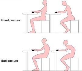 ©EDU
©EDU
Although elasticity is also a characteristic of skeletal tissue, if any muscle is held at a certain length (longer or shorter) for long periods of time, it may gradually alter its resting length.
Kyphosis is exaggerated curvature of the cervical vertebrae and can be due to the shoulder girdles being protracted forwards (hunched shoulders) for too long.
Over time this can stretch and lengthen the resting length of the muscles in the upper back. As these muscles are attached to the clavicle and the scapula, they can alter the position of the bones they are attached to.
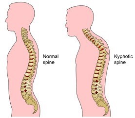 ©EDU
©EDU
Another common postural condition is lordosis, which is exaggerated curvature of the lumbar vertebrae. The lumbar vertebrae have a natural curve which helps this part of the spine to cope with the weight of the body, but if more weight is applied to the front of the body, for instance during pregnancy, or if the abdominal muscles are not utilised to help with everyday stabilisation, the lumbar curve can become exaggerated.
If this happens, the muscles surrounding the lumbar vertebrae (erector spinae, transversospinalis and segmental back muscles) become tighter and the opposing abdominal muscles may become longer and weaker.
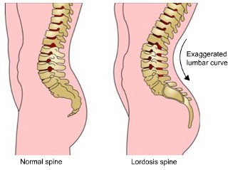 ©EDU
©EDU
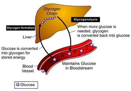
Muscles are a major site of glycogen storage in the human body. Glycogen is the storage form of glucose, which we use for energy. Glycogen is stored in the muscles and liver and converted into glucose whenever blood glucose levels begin to drop.
The average person stores 1500 – 2000 calories as glycogen and each gram of glycogen is stored with a few grams of water.
 ©EDU
©EDU
Diagram showing glucose molecules in the blood stream and a glycogen chain in the liver.
Glycogen formation: Glucose is converted into glycogen for stored energy.
Glycogenolysis: When more glucose is needed, glycogen is converted back into glucose.
This process maintains glucose levels in the bloodstream.
Thermogenesis means the creation of heat. Muscular contractions are thought to create as much as 85% of all body heat. Heat is a by-product of muscular contraction and this is utilised to maintain normal body temperature.
If body temperature drops, the central nervous system initiates an involuntary contraction of skeletal muscles known as shivering in order to generate heat and elevate the body temperature.
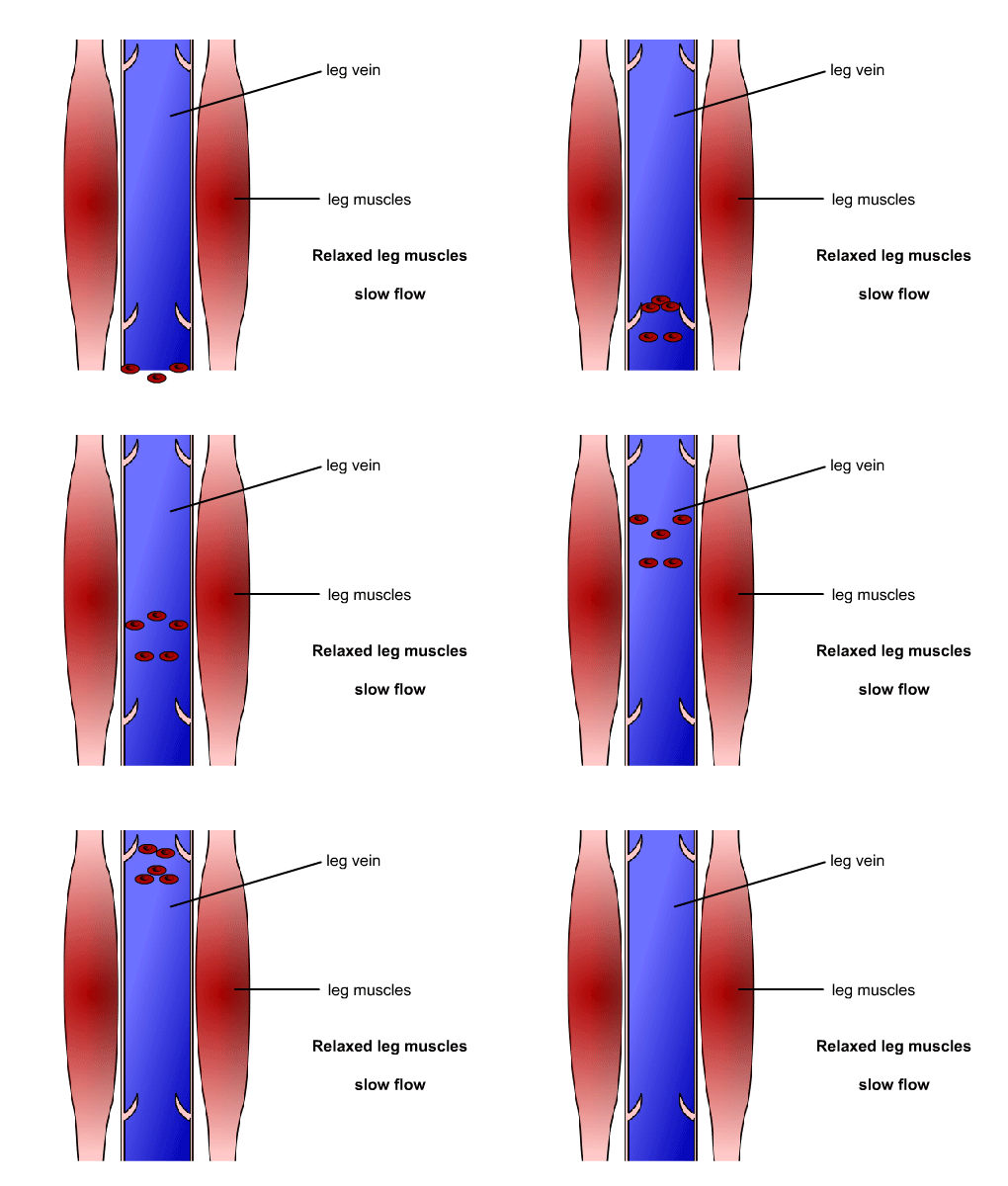
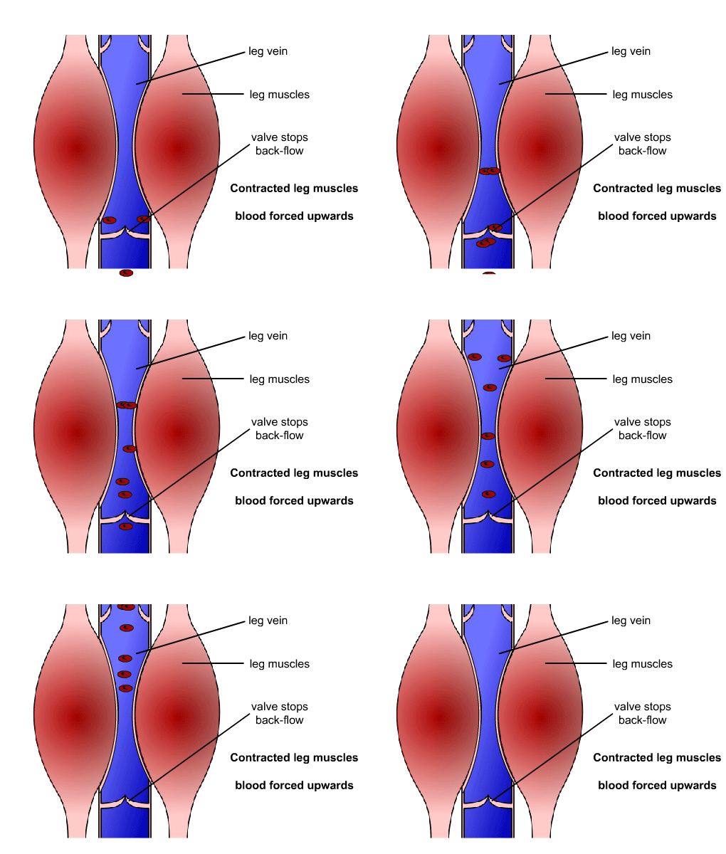
Although the walls of blood vessels do contain smooth muscle to help them contract and relax, the skeletal muscles also play a part in helping blood to return to the heart through the veins. This is particularly important in the lower limbs, where gravity makes venous return (blood flow back to the heart) more difficult.
When the skeletal muscles surrounding a vein contract, this gently squeezes the vein, and the flow of blood towards the heart is assisted by the pressure of the contracting muscle. This pressure also closes the lower valve in the vein, preventing deoxygenated blood being forced backwards.
 ©EDU
©EDU
 ©EDU
©EDU
Quiz
Muscles of the human body
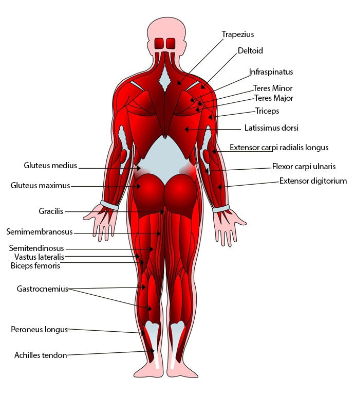
There are almost 700 skeletal muscles in the human body. Take a look at these diagrams and familiarise yourself with the major muscles shown.
Anterior muscles
Posterior muscles
UHI / CC0
Gluteus medius
Gluteus maximus
Gracilis
Semimembranosus
Semitendinosus
Vastus lateralis
Biceps femoris
Gastrocnemius
Peroneus longus
Achilles tendon
Trapezius
Deltoid
Infraspinatus
Teres Minor
Teres Major
Triceps
Latissimus dorsi
Extensor carpi radialis longus
Flexor carpi ulnaris
Extensor digitorium
Types of movement
As well as learning the names of the muscles, you need to know what each one does. Terms for the movements that muscles create are shown here.
|
Movement |
What happens |
Example |
|
Flexion |
The angle at a joint reduces |
Biceps muscle bending elbow |
|
Extension |
The angle at a joint increases |
Triceps muscle straightening the elbow |
|
Abduction |
Moving body parts away from one another or away from the body |
Lifting your arm or leg outwards |
|
Adduction |
Moving body parts closer together or towards the midline of the body |
Bringing the legs together |
|
Circumduction |
A body part rotates forming a cone shape or large circle |
Circling outstretched arms in a circle from the shoulder |
|
Rotation |
Moving a bone around its long axis medially or laterally |
Rotating (twisting) the thigh or arm towards the front midline of the body (medial rotation) or twisting away from the midline (lateral rotation) |
|
Elevation |
Lifting |
Lifting (shrugging) shoulders |
|
Depression |
Lowering |
Lowering shoulders |
|
Protraction |
Bringing forwards |
Moving shoulders forwards |
|
Retraction |
Bringing backwards |
Moving shoulders backwards |
Activity
Download this blank table in either Word or PDF and fill it in.
In the following pages, you will learn about the different muscles of the body. Use the Word document to take notes on these muscles. You may also want to note where each muscle is on the body or add an extra column at the end to include exercises for each muscle.
Luckily, many muscles hold a clue in their name as to where they are located, what they look like, or what they do in the body.
Read on to find out how to make remembering all these muscles a little easier!
Naming skeletal muscles:
Shape and location in the body
The names of skeletal muscles are often based on specific characteristics which may help you to remember some of their names. Muscles may be named based upon where they are located in the body, what shape they are, their size, how many origins (tendons) they have, the direction of muscle fibres or their action.
For example, the deltoid muscle of the shoulder is so-named after the Greek letter Delta, which is triangle-shaped. The trapezius in the upper back is so-named as it is shaped like a trapezium. The rhomboid muscles between the shoulder blades are rhomboid-shaped.
Delta
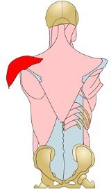
UHI / CC0
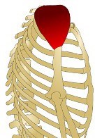
UHI / CC0
Trapezius
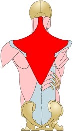
UHI / CC0
Rhomboid
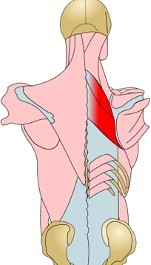
UHI / CC0
Muscle size or number of tendons
Muscles may be named depending upon their size:
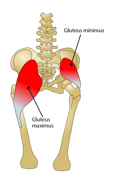
Gluteus minimus and maximus – the smallest and largest gluteal muscles.
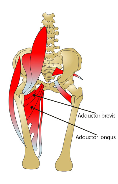
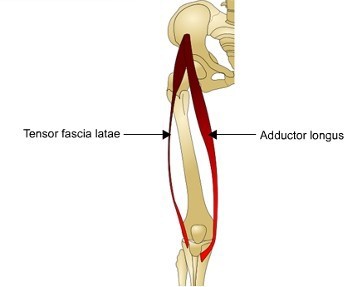
Adductor brevis and longus – the shortest and longest adductor muscles in the inner thigh.
There may also be a clue as to how many origins a muscle has in its name:
- The biceps muscle has two origins
- The triceps muscle has three origins
- The quadriceps muscles have four origins
Direction of muscle fibres
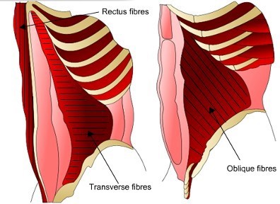
The direction of how muscle fibres are arranged may contribute to the name of the muscle, so knowing the name of these muscles might help you determine where the origin and insertion points are. All of the abdominal muscles are named according to the direction that the muscle fibres run in.
Transverse fibres run across the body E.g. Transverse abdominis.
Oblique fibres run diagonally to the midline E.g. Internal and external obliques.
Rectus fibres run parallel to the midline of the body E.g. Rectus abdominis.
The action of the muscle
There is often a connection between the muscle name and its action.
For example:
Adductor longus – the inner thigh muscle that adducts the leg towards the body
Tensor fascia latae – a band of connective tissue (fascia) which helps to create tension and rigidity down the outer thigh.
Major muscles of the upper body
Only the major skeletal muscles of the human body are included here with their main attachment points. It is recommended that you also use an anatomy textbook to familiarize yourself with smaller muscles and more specific attachment points.
Located at the back of the neck and posterior upper trunk, this muscle originates from the occipital bone, 7th cervical and all thoracic vertebrae and inserts into the clavicle and scapula.
It elevates the clavicle, adducts and rotates the scapula upwards, elevates and depresses the scapula and extends the head.
UHI / CC0
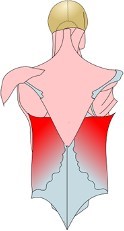
This large back muscle originates from the lower 6 thoracic and lumbar vertebrae, the crests of the sacrum and ilium and the lower 4 ribs.
It inserts into the humerus and extends, adducts and medially rotates the arm.
UHI / CC0
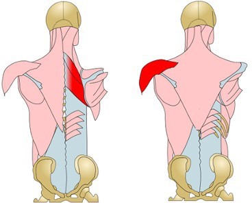
The deltoid (shoulder) muscle has three heads - the anterior (front), medial (middle), and posterior (rear) head.
It originates from the acromial extremity of the clavicle and inserts into the deltoid tuberosity of the humerus. The deltoid muscle abducts, flexes, extends, medially and laterally rotates the arm.
A greater number of muscle fibres are recruited from different parts of the deltoid (anterior, medial or posterior) for different movements and exercises.
Located between the shoulder blades, the rhomboids originate from the cervical and thoracic vertebrae and insert onto the scapulae. Both rhomboid muscles adduct the scapula (retraction), and also rotate them inferiorly.
UHI / CC0
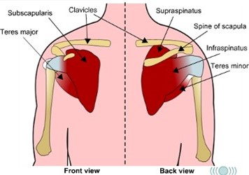
Located around the scapula and humerus, the subscapularis muscle together with the supraspinatus, infraspinatus and teres major/minor make up a small group of muscles known as the rotator cuff.
The subscapularis originates from the scapula and inserts into the lesser tubercle of the humerus. It medially rotates the arm.
The supraspinatus originates from the scapula and inserts into the lesser tubercle of the humerus. It assists the deltoid in arm abduction.
The infraspinatus originates from the scapula and inserts into the lesser tubercle of the humerus. It adducts and rotates the arm laterally.
The teres major and teres minor both originate on the scapula and insert into the humerus. The teres major assists in arm adduction and medial rotation, and also extends the arm; the teres minor rotates the arm laterally, extends and adducts it.
UHI / CC0
Front view:
- Clavicle
- Subscapularis
- Teres major
Back view:
- Clavicle
- Supraspinatus
- Spine of scapula
- Infraspinatus
- Teres mino
Quiz
A quick test of memory and muscle knowledge!
Put each muscle name to the correct box that corresponds with its description.
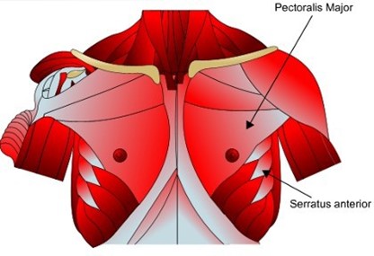
Pectoralis major, pectoralis minor and serratus anterior
The pectoralis major originates on the sternum and ribs and inserts into the humerus. It adducts the arms, bringing them together at the front of the body, a movement called lateral rotation.
The pectoralis minor originates on the superior 8 or 9 ribs and inserts into the scapula. It rotates the scapula upwards and outwards (protraction) and elevates the ribs if the scapula is fixed.
The serratus anterior originates from the clavicle, sternum and cartilage of the 2nd to the 6th ribs and inserts into the humerus. It flexes, adducts and medially rotates the arm, creating pushing and punching movements.
Biceps, brachialis and brachioradialis
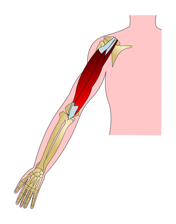
The bicep has two origins: the long head originates from the tubercle superior to the glenoid cavity (scapula); the short head originates from the coracoid process of the scapula. The biceps muscle inserts into the radial tuberosity on the radius and flexes the forearm at the elbow joint. Because it passes the shoulder joint, it also flexes the arm. The bicep also supinates the forearm, twisting the palm from facing the side of the body at rest to facing upwards.
The brachialis originates on the lower front of the humerus and inserts into the ulnar tuberosity and coronoid process of the ulna. It flexes the forearm (bends the elbow).
The brachioradialis originates on the middle and outer parts of the humerus and inserts into the radius. It flexes the forearm (bends the elbow).
UHI / CC0
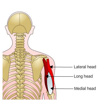
Triceps
The triceps muscle has three origins:
- the long head originates from above the glenoid cavity (scapula)
- the lateral head originates from the side and posterior of the humerus
- the medial head originates from the posterior humerus
The triceps insert into the olecranon of the ulna and extend (pull back) the arm (at the shoulder) and the forearm (at the elbow).
Erector Spinae and Quadratus Lumborum
Now let’s concentrate on the muscles of the mid-section or trunk.
The erector spinae are a group of back muscles consisting of the iliocostalis, longissimus and spinalis muscles. These muscles run lengthways along the spine and are responsible for extending (straightening) various regions of the spine and maintaining an erect posture. Most attach to the vertebrae, but some muscles originate or insert into the ribs or the iliac crest. Use your text book to familiarise yourself further with the erector spinae muscles.
The quadratus lumborum originates from the iliac crest and inserts into the 12th rib and first 4 lumbar vertebrae. Contraction of one side creates lateral flexion (side bending) of the vertebral column, and it also moves and fixes the 12th rib during breathing.
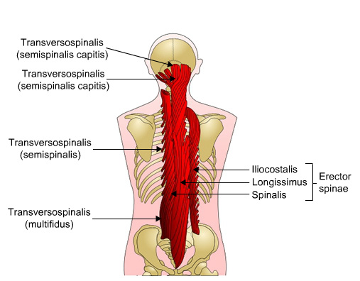
UHI / CC0
Transversospinalis (semispinalis capitis)
Transversospinalis (semispinalis)
Transversospinalis (multifidus)
Erector spinae:
- Iliocostalis
- Longissimus
- Spinalis
Abdominal muscles
The rectus abdominis originates on the pubic crest and pubic symphysis and inserts into the cartilage of the 5th to 7th ribs and xiphoid process. It flexes the vertebral column, aids in posture and helps create intra-abdominal pressure.
The transverse abdominus originates from the iliac crest, inguinal ligament, lumbar fascia and cartilage of lower 6 ribs. It inserts into the xiphoid process, the linea alba and the pubis and compresses the abdomen, also promoting good posture.
The internal oblique originates from the iliac crest and inguinal ligament and inserts into the cartilage of the lower 3 or 4 ribs and linea alba. Contraction of one side of the muscle creates lateral flexion or rotation of the spinal column.
Quiz
quick test of memory and muscle knowledge! Fill in each muscle name to the correct box that corresponds with its description.
Major muscles of the lower body
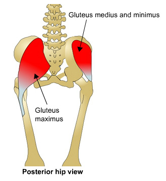
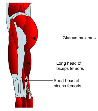
The gluteus maximus is the largest of the three gluteal muscles. It originates at the iliac crest, sacrum and coccyx and inserts into the femur. It extends and laterally rotates the thigh, moving the leg backwards and twisting it outwards away from the body.
The gluteus medius and minimus are much smaller and sit on the outside of the hip. They originate at the ilium and insert into the greater trochanter of the femur, abducting and rotating the thigh medially, moving the leg away from the body, or twisting it inwards.
The biceps femoris is the larger of the hamstring muscles at the back of the thigh. The long head originates from the ischial tuberosity, and the short head originates from the femur. It inserts into both the tibia and fibula, flexing the leg at the knee joint and extending the thigh.
The semitendinosus and semimembranosus
both originate at the ischial tuberosity and insert into different parts of the tibia. They both extend the thigh and flex the leg at the knee.
The psoas muscle originates at the lumbar vertebrae and inserts into the femur. It flexes and laterally rotates the thigh and also flexes the vertebral column. The iliacus originates at the iliac fossa and inserts into the tendon of the psoas. These two muscles are often called the ilio-psoas and treated as one muscle as they both have the same action.
The rectus femoris originates from the iliac spine and inserts into the tibial tuberosity via the patella tendon. It extends the leg at the knee joint and flexes it at the thigh.
The vastus lateralis, medialis and intermedius originate on the femur and insert into the tibial tuberosity via the patella tendon. The names of the muscles denote their position on the thigh, with the vastus lateralis on the outside, the vastus medialis innermost and the intermedius in the middle. All vastus muscles extend the leg at the knee joint. These muscles and the rectus femoris are the quadriceps.
The sartorius is the longest muscle in the body. It originates from the iliac spine and inserts into the tibia. It flexes the leg at the hip and also controls lateral rotation.
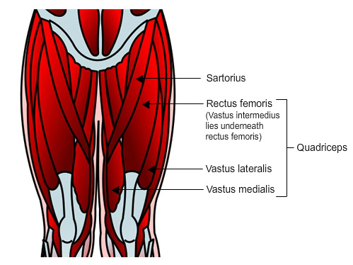
UHI / CC0
Sartorius
Quadriceps:
- Rectus femoris (Vastus intermeduis lies underneath rectus femoris)
- Vastus lateralis
- Vastus medialis
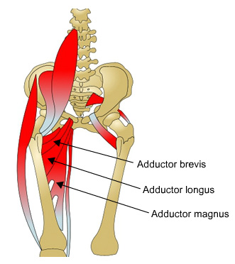
There are three adductor muscles, the adductor longus, adductor brevis, and adductor magnus. The longest adductor muscle is the adductor longus. It originates from the pubic crest and pubic symphysis and inserts into the femur. It adducts, flexes and medially rotates the thigh.
The adductor brevis is the shortest adductor. It originates from the pubis and inserts into the femur, and adducts, flexes and medially rotates the thigh.
The largest adductor magnus muscle originates from the pubis and ishium and inserts into the femur. It adducts and medially rotates the thigh. The anterior part of this muscle flexes the thigh; the posterior fibres aid in thigh extension.
The pectineus originates from the pubis and inserting into the femur, and flexes and adducts the thigh.
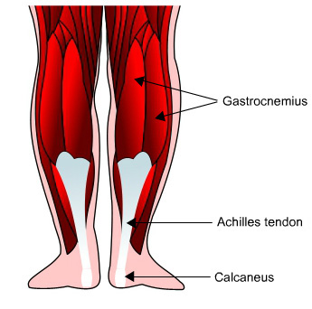
The gastrocnemius originates from the lateral and medial condyles of the femur and the knee capsule and inserts into the calcaneus via the Achilles tendon. It plantar flexes the foot and aids in knee flexion, allowing us to point our toes and bend the knee.
The soleus originates from the fibula and tibia and also inserts into the calcaneus via the Achilles tendon. It plantar flexes the foot (points the toes).
The tibialis anterior is located on the shin, originating on the tibia and inserting into the first metatarsal and first cuneiform. It dorsiflexes and inverts the foot.
The peroneus longus originates from the fibula and tibia and inserts into the first metatarsal and first cuneiform. The peroneus brevis originates from the fibula and inserts into the 5th metatarsal. Both muscles plantar flex and evert the foot.
Quiz
How muscles work
Muscles are attached to bones at their origin and insertion points by tendons. Tendons are made of collagenous fibres and are exceptionally strong yet pliable.
They are a continuation of the connective tissue that surrounds each muscle fibre and bundle of fibres.
The tendon attaches the muscle to two (or more) points. The first is the point of origin, which does not usually move when the muscle contracts; the second attachment is the point of insertion, which moves when the muscle contracts or relaxes.

UHI / CC0
(Click image to toggle animation on/off)
Muscle is attached to bone by tendons, ligament attaches two bones at the joint, joint is protected by cartilage in a joint capsule.
Muscles work in pairs – reciprocal relaxation
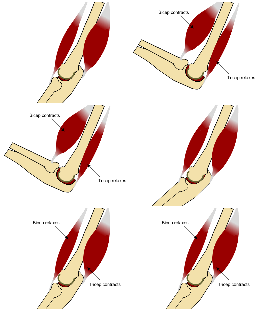
When a prime mover contracts, the opposing antagonist muscle relaxes. This is called reciprocal relaxation. Consider the example of flexing the arm by bending the elbow. This is achieved by contraction of the biceps muscle which raises the radius. At the same time, the triceps muscle (the antagonist) relaxes.
Other muscles which cross the elbow joint anteriorly may act as synergists (brachialis, for example) and during this movement, the deltoid muscles are fixating to keep the upper arm still.
The roles of the biceps and triceps are reversed when the forearm is extended (straightened) against resistance. Here the triceps muscle becomes the prime mover and the biceps muscle the antagonist as it relaxes.
Sensory receptors
Proprioceptors are sensory receptors in muscles, tendons and joints that provide information about muscle or tendon tension and movement.
Muscle spindles are specialised groups of muscle fibre which monitor changes in muscle length. They do not contain contractile proteins, but respond to nervous impulses and contract in response to a sudden or prolonged stretch.
When a muscle is stretched, a message is sent to the nervous system, resulting in the muscle contracting to prevent over-stretching, hence the tightness felt when a muscle is stretched. If the stretch is controlled and muscle length remains the same for approximately 30 seconds, the proprioceptors will send a second message resulting in relaxation of the muscle and enabling enhanced flexibility training.
Golgi tendon organs are capsules of connective tissue containing sensory fibres located at the musclo-tendon junction. They monitor the force of muscular contraction and tendon tension. Under increased tension, nervous impulses are sent to the nervous system to help protect tendons and muscles from damage.
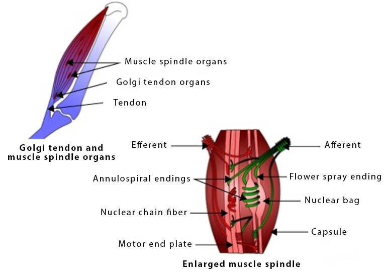
UHI /CC0
Golgi tendon and muscle spindle organs:
- Muscle spindle organs
- Golgi tendon organs
- Tendon
Enlarged muscle spindle:
- Efferent
- Afferent
- Annulospiral endings
- Flower spray endings
- Nuclear chain fibre
- Nuclear bag
- Motor end plate
- Capsule
Types of muscular contraction
There are four types of muscular contraction: Concentric, Eccentric , Isometric , and Isokinetic.
The concentric and eccentric contractions together are called an isotonic contraction.
When an opposing muscle relaxes and lengthens to enable the agonist muscle to contract, there is no tension in the antagonistic muscle. This is called reciprocal relaxation.
During a concentric muscular contraction, the angle at the joint decreases as the origin and insertion points of the muscle come closer together. The muscle shortens and develops tension. An example of this is in the biceps muscle as the arm bends.

UHI / CC0
(Click image to toggle animation on/off)
Muscle is attached to bone by tendons, ligament attaches two bones at the joint, joint is protected by cartilage in a joint capsule.
During an eccentric contraction, the angle at the joint increases as the origin and insertion points move further apart and the muscle lengthens as it develops tension. Note that the eccentric contraction is usually in the same muscle as the concentric contraction, in the biceps muscle, for example, when a heavy weight is being lowered.
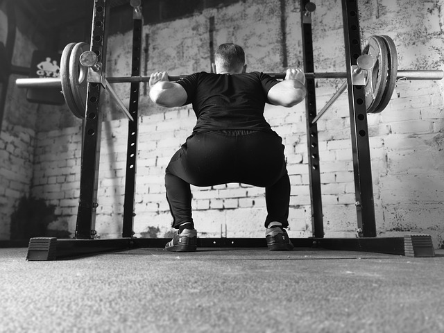
In an isometric contraction, there is still tension in the muscle, but the muscle remains the same length, for example when pushing against a wall, or during exercises such as a squat hold, where a position is held for a period of time. Stabilising muscles often contract in this way to help maintain body positioning whilst other muscles bring about a movement in a different body part.
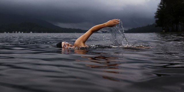
Isokinetic contractions take place on specialised exercise machines which provide an equal resistance throughout the movement. During most exercises, we experience a more difficult section of the movement, usually when the force of gravity is at its greatest, but in water, or on specialised exercise equipment, the force against the muscle remains the same and so the speed of muscular contraction also remains the same.
The structure of skeletal muscle
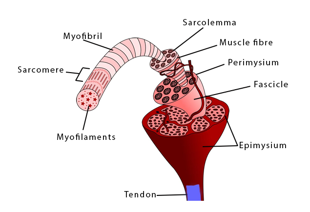
Muscles are made up of long, cylindrical muscle fibres. Each muscle fibre contains several nuclei and is surrounded by a thin membrane called a sarcolemma. Blood capillaries surround the sarcolemma so that when the muscle contracts it is ensured of a supply of oxygenated, nutrient-rich blood. Thin strands called myofibrils make up each muscle fibre, and these contain contractile protein filaments called actin and myosin.
Muscle fibres are arranged within the muscle in bundles called fascicles. The muscle fibres run from one end of the fascicle to the other. Each bundle (fascicle) is surrounded by a protective connective tissue called the perimysium. The fibres and fascicles all run parallel to one another, providing greater strength.
The muscle is surrounded by a layer of connective tissue called the epimysium.
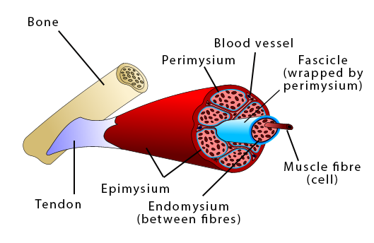
UHI / CC0
Bone
Perimysium
Blood vessel
Fascicle (wrapped by perimysium)
Muscle fibre (cell)
Endomysium (between fibres)
Epimysium
Tendon
UHI / CC0
Myofilaments
Sarcomere
Myofibril
Sacrolemma
Muscle fibre
Perimysium
Fascicle
Epimysium
Tendon
Structure of skeletal muscle explained in simple terms (YouTube 2:10)
Sliding filament theory
Each myofibril is divided into sections called sarcomeres which contain the protein filaments (myofilaments) actin and myosin.
Muscular movement occurs as a result of the sliding action of the protein filaments (myofilaments) actin and myosin. The end of each sarcomere is divided by a Z line, and when actin and myosin slide past one another this causes the sarcomere to contract and shorten, and the Z lines move closer together.
As each sarcomere shortens along the myofibril, the entire muscle fibre shortens, and hence the muscle contracts. When the muscles relax, the sarcomere lengthens again.
Relaxed muscle

Partially contracted muscle

The actin filaments slide over the myosin filaments and the Z lines get closer together.
Fully contracted muscle

The actin filaments slide over the myosin filaments and the Z lines get even closer.
UHI / CC0
A sarcomere is the functional unit of a myofibril and goes from Z line to Z line. Each sarcomere contains a number of light or dark coloured bands, dependant upon whether each section contains only thin strands of actin, only thick bands of myosin, or both.
The H zone contains thick but not thin filaments
The A band contains both types of filament
The I band appears lighter (thin actin filaments)
The I band goes from the end of the thick myosin filaments, across to the neighbouring sarcomere and ends at the beginning of the next group of myosin filaments – this is the lighter part of the myofibril which excludes myosin filaments.
Myosin and actin give the myofibril a striped appearance under a microscope as the myosin, which is thicker, produces a darker colour than the thinner actin filament, particularly where the two filaments overlap.
Quiz
Have a go at labelling the structure of the skeletal muscle.
How muscles and the nervous system work together
Contraction of skeletal muscles is stimulated by nerve impulses from the nervous system. The nerve fibres which supply nervous impulses to the muscles are called motor neurones. Electrical impulses transmitted through the motor neurones cause muscular contraction. The motor neurone together with all the muscle fibres it stimulates is called the motor unit.
One nerve fibre (motor neurone) makes contact with a number of muscle fibres. The number of muscle fibres in a motor unit depends upon the type of work being done and the amount of control needed: muscles controlling precise movements such as those in the eye have many small motor units, but bigger movements in large muscles such as the leg are a result of hundreds of fibres responding to a single motor unit. The greater the larger the number of motor units engaged, the stronger the muscular contraction.
UHI / CC0
- Sarcolemma
- Motor neurone
- Neurotransmitter released
- Axon terminal
- Mitochondrium
- Sarcomere
- Motor end plate
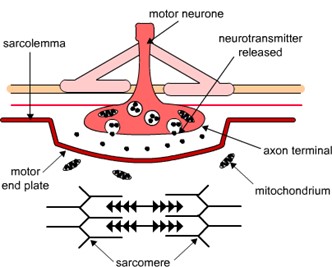
Each motor neurone has a threadlike axon that extends from the spinal cord to a group of muscle fibres. The end of each motor neurone divides into many branches and may stimulate over 100 neighbouring muscle fibres.
The sarcolemma (membrane) surrounding each muscle fibre is pierced by a single branch of the motor neurone known as an axon terminal. The area of the sarcolemma where the branch of the nerve ends is called the motor end plate. The motor end plate and the axon terminal together form the neuromuscular junction.
At the end of each axon terminal is a cluster of synaptic end bulbs containing sacs filled with neurotransmitter molecules.
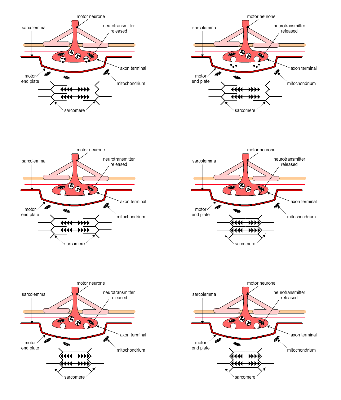
The neuromuscular junction reacts to an impulse called a nerve action potential from the motor neurone (nerve cell). Because there is a small gap between the axon terminal in the nerve ending and the motor end plate in the muscle fibre, a neurotransmitter is released from the synaptic end bulbs, which travels across the gap. Acetylcholine (Ach) is the neurotransmitter released at the neuromuscular junction.
Diffusion of acetylcholine into the sarcolemma (the membrane of the muscle fibre) starts a sequence of chemical reactions with calcium and sodium ions. These produce something called an action potential in the muscle fibre. Each sarcomere, the functional unit of a myofibril contains two transverse tubules which are folds in the sarcolemma of the muscle fibre filled with extracellular fluid. It is along these T-tubules that the muscle action potential travels, opening channels that release calcium ions as it travels along each sarcomere.
UHI / CC0
(Click image to toggle animation on/off)
Muscular contraction
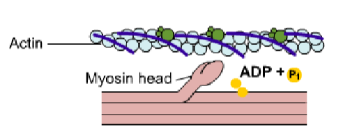
A nervous impulse to the sarcomere prompts the release of calcium ions from the sarcoplasmic reticulum into the sarcoplasm, coming into contact with the actin myofilaments.
The calcium ions bind to a protein (troponin) on the thin strands of actin, and the troponin-tropomyosin complex changes shape and exposes sites on the actin filament.
These exposed sites form cross bridges with the heads of the myosin filament and movement is initiated.
Energy is required to move and release the head of the myosin filament. The calcium ion stimulates energy to be released from an energy substrate in the muscle cell called adenosine triphosphate (ATP).
The release of energy and the movement of the myosin heads pull the actin filament towards the thicker myosin filament. At any one instant during muscular contraction, approximately half of the myosin heads are bound to actin, inducing the power stroke that creates this contraction.
At the end of the movement the myosin head is released with energy from more ATP, and the cycle is repeated, possibly over 100 times a second. As the actin myofilaments are attached to the Z-lines of the sarcomere, the length of the sarcomere shortens and the whole muscle contracts.
Muscles relax when the nervous impulse stops and calcium ions leave the actin filaments and return to storage in the sarcomere.
- Actin
- Myosin head
- ADP + P1
Action potential causes depolarization and release of CA2+ which attaches to the troponin on the actin filament
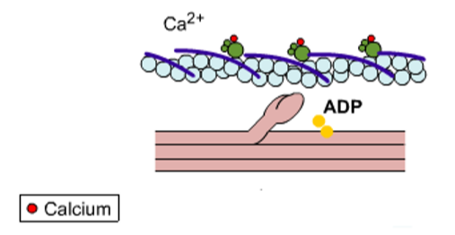
- Calcium
- CA2+
- ADP
Myosin pulls actin strand inwards, towards the centre of the sarcomere, and the Z lines get closer together.
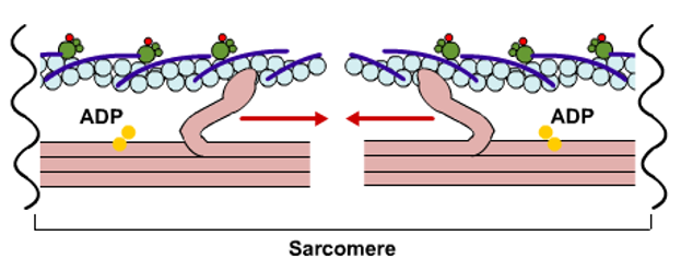
- ADP
- Sarcomere
- Calcium
UHI / CC0
The all or nothing theory
When we use a muscle, we estimate how much force is needed and recruit a number of muscle fibres to contract via our nerve fibres. This is rarely all of the muscles fibres in a muscle, but every fibre responding to the ‘fired’ motor neurone contracts fully. This is the all or nothing theory.
Muscle fibres either contract fully if stimulated by the motor neurone they are connected to or are not stimulated at all if the motor neurone has not been recruited for a movement to take place. Muscles fibres which have not been stimulated by a motor neurone will shorten and lengthen passively as the muscle changes its length. The entire muscle shortens, but depending on the amount of force required, some motor units will be fully contracted while others remain relaxed.
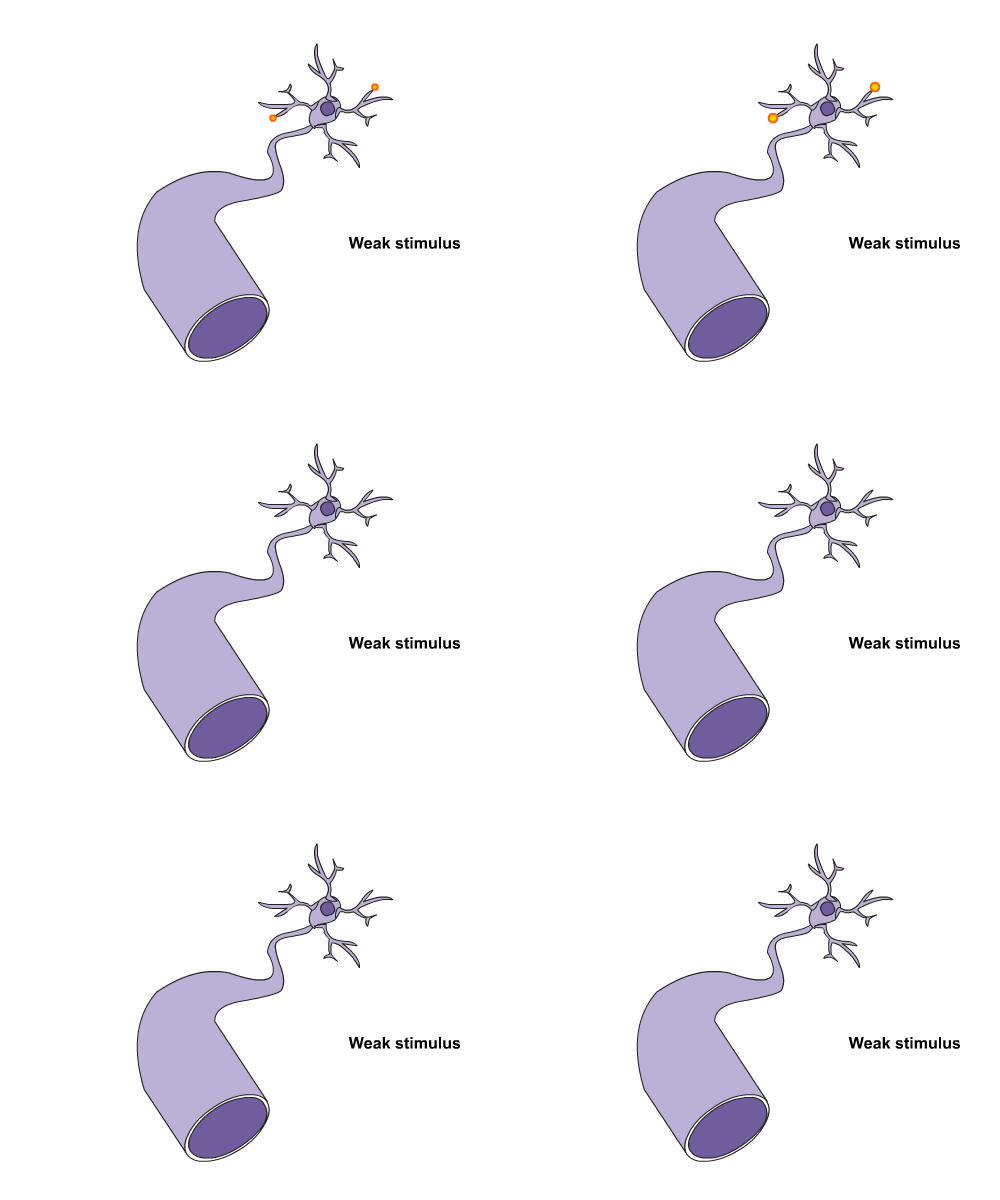
UHI / CC0
(Click image to toggle animation on/off)
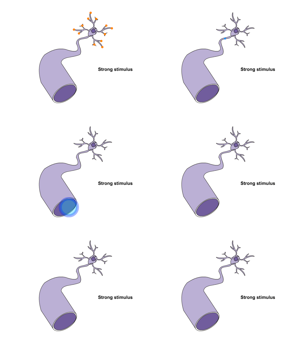
UHI / CC0
(Click image to toggle animation on/off)
Quiz
Before you go on, have a go at completing this paragraph to test your knowledge on how muscles and the nervous system work together by matching the words to the correct areas.
Test your knowledge on the muscular system
Now it’s time to test your knowledge of the muscular system. There are 10 multiple choice questions to complete. You might want to take some time to read your notes, draw diagrams or watch the animations again before you test yourself.
Summary
You have completed your study of the muscular system.
You should now have a good knowledge and understanding of the anatomy and physiology of the muscular system.
You should be able to...
- Describe the characteristics of, and differences between different types of muscle.
- Discuss the various functions of skeletal muscle.
- Describe the anatomy of muscle and muscle fibres.
- Know the location, insertion and origin points, and movements of the major muscles in the body.
- Understand how muscular contraction occurs and be able to describe it.
If you think that your knowledge or understanding of any section of the muscular system could be improved further, go back to the relevant sections and work through them again, taking time to make notes and complete the activities.
 ©EDU
©EDU
